File:2012 Sombke et al f05.png
Size of this preview: 544 × 599 pixels. Other resolutions: 218 × 240 pixels | 436 × 480 pixels | 697 × 768 pixels | 930 × 1,024 pixels | 2,001 × 2,204 pixels.
Original file (2,001 × 2,204 pixels, file size: 6.11 MB, MIME type: image/png)
Captions
Captions
Add a one-line explanation of what this file represents
Summary edit
| Description |
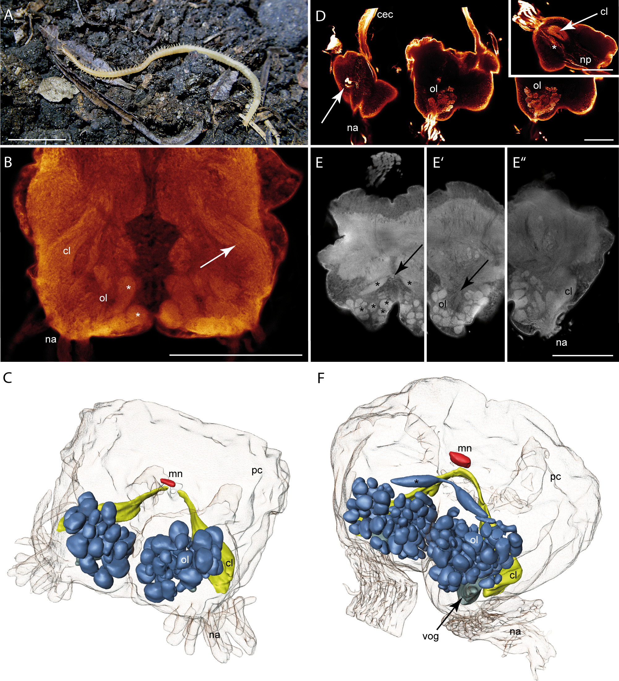
English: Figure 5 Geophilomorpha. A Geophilus carpophagus. B Single horizontal optical section (cLSM) of an autofluorescence preparation of the brain of Haplophilus subterraneus showing olfactory glomeruli (asterisks) and the structural composition of the corpus lamellosum (arrow). C 3D reconstruction
of the brain of H. subterraneus with deutocerebral neuropils and midline neuropil. Blue = olfactory glomeruli, yellow = corpus lamellosum, red = midline neuropil. Contralateral connection of the CL is not shown. D Single horizontal optical sections (cLSM) of a neurobiotin backfill of the right antennal nerve in Stigmatogaster dimidiatus from ventral to dorsal. Left: several somata stained by the neurobiotin backfill (arrow) and neurite projections into a circumesophageal connective. Middle: Antennal nerves and olfactory glomeruli. Right: The slightly concave appearance of the olfactory lobe. Inset: Sagittal optical section of the same preparation showing the structural composition of the corpus lamellosum (arrow) and neurite projections (asterisk). E Single horizontal optical sections (cLSM) of an autofluorescence preparation of the brain of S. dimidiatus from dorsal to ventral. Olfactory glomeruli (asterisks) and the contralateral connection between the posteroventral OG (arrow). E’ Concave appearance of the olfactory lobe (arrow). E’’ ventrolateral position of the corpus lamellosum. F 3D reconstruction of the brain of S. dimidiatus with deutocerebral neuropils and midline neuropil. Blue = olfactory glomeruli, gray = bigger ventral olfactory glomerulus, yellow = corpus lamellosum, red = midline neuropil. Abbreviations: cec circumesophageal connective, cl corpus lamellosum, mn midline neuropil, na nervus antennalis, np neurite projections, ol olfactory lobe, pc protocerebrum, vog ventral olfactory glomerulus. Scalebars: A = 5 mm, B, D, E = 100 μm. |
| Date | |
| Source | Sombke, Andy; Lipke, Elisabeth; Kenning, Matthes; Müller, Carsten HG; Hansson, Bill S.; Harzsch, Steffen (2012-01-03). Comparative analysis of deutocerebral neuropils in Chilopoda (Myriapoda): implications for the evolution of the arthropod olfactory system and support for the Mandibulata concept. BMC Neuroscience 13 (1): 1. doi:10.1186/1471-2202-13-1 |
| Author | Andy Sombke, Elisabeth Lipke, Matthes Kenning, Carsten HG Müller, Bill S Hansson, Steffen Harzsch |
Licensing edit
This file is licensed under the Creative Commons Attribution 2.0 Generic license.
- You are free:
- to share – to copy, distribute and transmit the work
- to remix – to adapt the work
- Under the following conditions:
- attribution – You must give appropriate credit, provide a link to the license, and indicate if changes were made. You may do so in any reasonable manner, but not in any way that suggests the licensor endorses you or your use.
File history
Click on a date/time to view the file as it appeared at that time.
| Date/Time | Thumbnail | Dimensions | User | Comment | |
|---|---|---|---|---|---|
| current | 01:15, 2 September 2022 | | 2,001 × 2,204 (6.11 MB) | Junnn11 (talk | contribs) | Uploaded a work by Andy Sombke, Elisabeth Lipke, Matthes Kenning, Carsten HG Müller, Bill S Hansson, Steffen Harzsch from Sombke, Andy; Lipke, Elisabeth; Kenning, Matthes; Müller, Carsten HG; Hansson, Bill S.; Harzsch, Steffen (2012-01-03). [https://www.researchgate.net/publication/51980717 Comparative analysis of deutocerebral neuropils in Chilopoda (Myriapoda): implications for the evolution of the arthropod olfactory system and support for the Mandibulata concept]. BMC Neuroscience 13 (1):... |
You cannot overwrite this file.
File usage on Commons
There are no pages that use this file.