File:24-Color 3D FISH Representation and Classification of Chromosomes in a Human G0 Fibroblast Nucleus 10.1371 journal.pbio.0030157.g001-M.jpg
24-Color_3D_FISH_Representation_and_Classification_of_Chromosomes_in_a_Human_G0_Fibroblast_Nucleus_10.1371_journal.pbio.0030157.g001-M.jpg (575 × 600 pixels, file size: 77 KB, MIME type: image/jpeg)
Captions
Captions
| Description24-Color 3D FISH Representation and Classification of Chromosomes in a Human G0 Fibroblast Nucleus 10.1371 journal.pbio.0030157.g001-M.jpg |
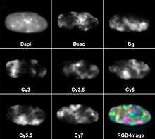
English: Color 3D FISH Representation and Classification of Chromosomes in a Human G0 Fibroblast Nucleus
(A) A deconvoluted mid-plane nuclear section recorded by wide-field microscopy in eight channels: one channel for DAPI (DNA counterstain) and seven channels for the following fluorochromes: diethylaminocoumarin (Deac), Spectrum Green (SG), and the cyanine dyes Cy3, Cy3.5, Cy5, Cy5.5, and Cy7. Each channel represents the painting of a CT subset with the respective fluorochrome. The combinatorial labeling scheme is described in Materials and Methods. RGB images of the 24 differently labeled chromosome types (1–22, X, and Y) were produced by superposition of the seven channels (bottom right). (B) False color representation of all CTs visible in this mid-section after classification with the program goldFISH. (C) 3D reconstruction of the complete CT arrangement in the nucleus viewed from different angles. (D) Simulation of a human fibroblast model nucleus according to the SCD model (see Materials and Methods). The first image shows 46 statistically placed rods representing the 46 human chromatids. The next three images simulate the decondensation process and show the resulting CT arrangement obtained after different numbers of Monte Carlo relaxation steps (200, 1,000, and 400,000). This set of figures is taken from Video S1. |
||
| Date | |||
| Source | Bolzer et al., (2005) Three-Dimensional Maps of All Chromosomes in Human Male Fibroblast Nuclei and Prometaphase Rosettes. PLoS Biol 3(5): e157 DOI: 10.1371/journal.pbio.0030157, part of Figure 1. | ||
| Author | Andreas Bolzer, Gregor Kreth, Irina Solovei, Daniela Koehler, Kaan Saracoglu, Christine Fauth, Stefan Müller, Roland Eils, Christoph Cremer, Michael R. Speicher, Thomas Cremer | ||
| Permission (Reusing this file) |
|
||
| Other versions |
  |
File history
Click on a date/time to view the file as it appeared at that time.
| Date/Time | Thumbnail | Dimensions | User | Comment | |
|---|---|---|---|---|---|
| current | 22:59, 15 November 2006 |  | 575 × 600 (77 KB) | Patho (talk | contribs) | {{Information |Description= DOI: 10.1371/journal.pbio.0030157.g001 View larger image Download source file Help Figure 1.24-Color 3D FISH Representation and Classification of Chromosomes in a Human G0 Fibroblast Nucleus (A) A deconvoluted mid-plane nucle |
You cannot overwrite this file.
File usage on Commons
The following 2 pages use this file:
File usage on other wikis
The following other wikis use this file:
- Usage on de.wikibooks.org
- Usage on sh.wikipedia.org
- Usage on sr.wikipedia.org
Metadata
This file contains additional information such as Exif metadata which may have been added by the digital camera, scanner, or software program used to create or digitize it. If the file has been modified from its original state, some details such as the timestamp may not fully reflect those of the original file. The timestamp is only as accurate as the clock in the camera, and it may be completely wrong.
| JPEG file comment | Handmade Software, Inc. Image Alchemy v1.10.2d22 |
|---|

