File:3D model of the deep cerebellar nuclei.png

Size of this preview: 800 × 429 pixels. Other resolutions: 320 × 172 pixels | 640 × 344 pixels | 1,024 × 550 pixels | 1,872 × 1,005 pixels.
Original file (1,872 × 1,005 pixels, file size: 472 KB, MIME type: image/png)
Captions
Captions
Add a one-line explanation of what this file represents
Summary
edit
| Description |
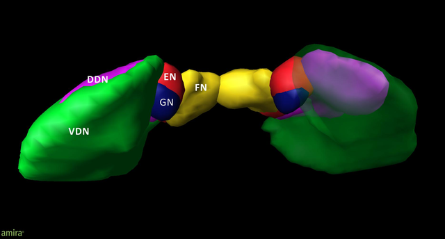
English: 3D model of the deep cerebellar nuclei (posterior to anterior view) of an individual brain (post mortem brain10); visualization by Amira 5.6.0 (www.amira.com). Dorsal dentate nucleus (DDN; magenta); ventral dentate nucleus (VDN; green); emboliform nucleus (EN; red); globose nucleus (GN; blue); fastigial nucleus (FN; yellow). Due to the smoothing, the dentate appears less denticulated than it is. The transparency of the right ventral dentate nucleus clarifies the partly covered extend of the DDN. |
| Date | Published online: 13 May 2015 |
| Source | Tellmann S, Bludau S, Eickhoff S, Mohlberg H, Minnerop M and Amunts K (2015) Cytoarchitectonic mapping of the human brain cerebellar nuclei in stereotaxic space and delineation of their co-activation patterns. Front. Neuroanat. 9:54. https://doi.org/10.3389/fnana.2015.00054 |
| Author | Stefanie Tellmann, Sebastian Bludau, Simon Eickhoff, Hartmut Mohlberg, Martina Minnerop and Katrin Amunts |
Licensing
edit
This file is licensed under the Creative Commons Attribution 4.0 International license.
- You are free:
- to share – to copy, distribute and transmit the work
- to remix – to adapt the work
- Under the following conditions:
- attribution – You must give appropriate credit, provide a link to the license, and indicate if changes were made. You may do so in any reasonable manner, but not in any way that suggests the licensor endorses you or your use.
File history
Click on a date/time to view the file as it appeared at that time.
| Date/Time | Thumbnail | Dimensions | User | Comment | |
|---|---|---|---|---|---|
| current | 04:24, 2 June 2019 | | 1,872 × 1,005 (472 KB) | Was a bee (talk | contribs) | {{Information |Description={{en|1=3D model of the deep cerebellar nuclei (posterior to anterior view) of an individual brain (post mortem brain10); visualization by Amira 5.6.0 (www.amira.com). Dorsal dentate nucleus (DDN; magenta); ventral dentate nucleus (VDN; green); emboliform nucleus (EN; red); globose nucleus (GN; blue); fastigial nucleus (FN; yellow). Due to the smoothing, the dentate appears less denticulated than it is. The transparency of the right ventral dentate nucleus clarifies... |
You cannot overwrite this file.
File usage on Commons
There are no pages that use this file.