File:3 10 helix neg49 neg26 sideview.png

Size of this preview: 450 × 599 pixels. Other resolutions: 180 × 240 pixels | 360 × 480 pixels | 894 × 1,191 pixels.
Original file (894 × 1,191 pixels, file size: 156 KB, MIME type: image/png)
Captions
Captions
Add a one-line explanation of what this file represents
Summary
edit
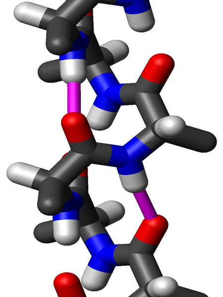
| Description | Close-up sideview of a "stick" model of an 310 helix of poly-alanine using the dihedral angles φ=-49° and ψ=-26° and the Engh&Huber bond geometry. Two hydrogen bonds to the same peptide group are highlighted in magenta; the O-H distance is 1.83 Å (183 pm). The PDB file was made by me on 18 October 2006 using my own software and visualized by me using MOLMOL. I release this image under the GFDL. |
| Date | 18 October 2006 (original upload date) |
| Source | No machine-readable source provided. Own work assumed (based on copyright claims). |
| Author | No machine-readable author provided. WillowW assumed (based on copyright claims). |
Licensing
edit
I, the copyright holder of this work, hereby publish it under the following license:

|
Permission is granted to copy, distribute and/or modify this document under the terms of the GNU Free Documentation License, Version 1.2 or any later version published by the Free Software Foundation; with no Invariant Sections, no Front-Cover Texts, and no Back-Cover Texts. A copy of the license is included in the section entitled GNU Free Documentation License. |
| This file is licensed under the Creative Commons Attribution-Share Alike 3.0 Unported license. | ||
| ||
| This licensing tag was added to this file as part of the GFDL licensing update. |
File history
Click on a date/time to view the file as it appeared at that time.
| Date/Time | Thumbnail | Dimensions | User | Comment | |
|---|---|---|---|---|---|
| current | 15:27, 18 October 2006 | | 894 × 1,191 (156 KB) | WillowW (talk | contribs) | Close-up sideview of a "stick" model of an 3<sub>10</sub> helix of poly-alanine using the dihedral angles φ=-49° and ψ=-26° and the Engh&Huber bond geometry. Two hydrogen bonds to the same peptide group are highlighted in magenta; the O-H distance is |
You cannot overwrite this file.
File usage on Commons
There are no pages that use this file.
File usage on other wikis
The following other wikis use this file:
- Usage on en.wikipedia.org
- Usage on es.wikipedia.org
- Usage on fr.wikipedia.org
- Usage on ja.wikipedia.org
- Usage on mk.wikipedia.org
- Usage on ru.wikipedia.org
- Usage on sh.wikipedia.org
- Usage on sr.wikipedia.org
- Usage on zh.wikipedia.org