File:41467 2016 Article BFncomms10543 Fig1 HTML.webp

Size of this PNG preview of this WEBP file: 514 × 600 pixels. Other resolutions: 206 × 240 pixels | 411 × 480 pixels | 946 × 1,104 pixels.
Original file (946 × 1,104 pixels, file size: 81 KB, MIME type: image/webp)
File information
Structured data
Captions
Captions
Add a one-line explanation of what this file represents
Summary edit
| Description41467 2016 Article BFncomms10543 Fig1 HTML.webp |
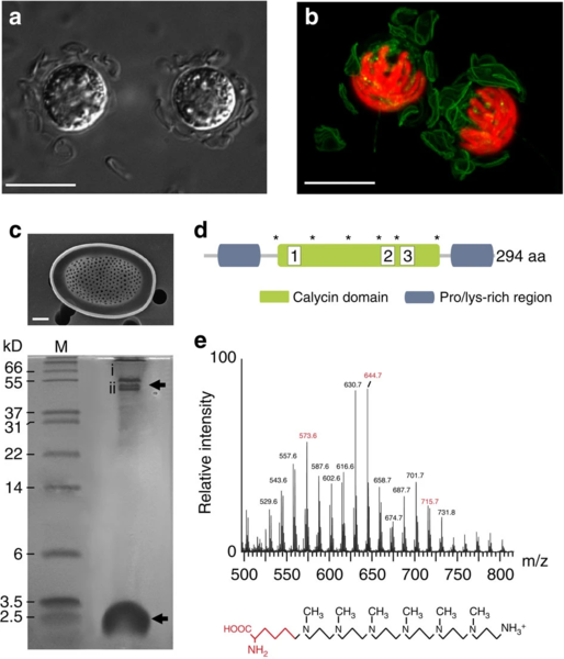
English: (a) Differential interference contrast (DIC) microscopy image of Prymnesium neolepis cells displaying the loose covering of silica scales. Scale bar, 10 μm. (b) Confocal microscopy of a P. neolepis cell showing incorporation of the fluorescent dye HCK-123 into newly formed silica scales (green). Chlorophyll autofluorescence is shown in red. The 3D-projection was generated from compiling a Z-stack of 15 images. Scale bar, 10 μm. (c) Tricine/SDS–PAGE of organic components released after dissolution of silica scales with NH4F. A SEM image of an isolated silica scale is also shown (Scale bar, 1 μm). The higher molecular weight component around 50 kDa is a single protein that runs as two bands (i, ii), whereas the low-molecular-weight components around 2.5 kDa are long-chain polyamines (LCPA). M, molecular-weight markers. (d) Domain organization of the lipocalin-like protein (LPCL1) identified from both protein bands in NH4F extracted silica scales. The approximate positions of the proline/lysine-rich regions and the calycin domain (IPR012674) are shown. Also shown are the positions of six highly conserved cysteines (asterisk) that may be involved in the formation of disulphide bridges. (e) Long-chain polyamines (LCPAs) from P. neolepis silica scales. Electrospray ionization mass spectrometry (ESI-MS) of the low-molecular-weight NH4F-soluble fraction of silica scales revealed a series of mass peaks separated by 71 Da (highlighted in red), characteristic of N-methyl propyleneimine units. The additional mass peaks ±14 Da may indicate different methylation states, as is commonly observed in LCPAs. The proposed structure of the LCPAs in P. neolepis is shown with the putative lysine residue is highlighted in red. |
|
| Date | ||
| Source |
Fig. 1 at https://www.nature.com/articles/ncomms10543 A role for diatom-like silicon transporters in calcifying coccolithophores In: Nat Commun 7, 10543 (2016). doi:10.1038/ncomms10543 |
|
| Author | Grażyna M. Durak, Alison R. Taylor, Charlotte E. Walker, Ian Probert, Colomban de Vargas, Stephane Audic, Declan Schroeder, Colin Brownlee, Glen L. Wheeler | |
| Other versions |
|
Licensing edit
This file is licensed under the Creative Commons Attribution-Share Alike 4.0 International license.
- You are free:
- to share – to copy, distribute and transmit the work
- to remix – to adapt the work
- Under the following conditions:
- attribution – You must give appropriate credit, provide a link to the license, and indicate if changes were made. You may do so in any reasonable manner, but not in any way that suggests the licensor endorses you or your use.
- share alike – If you remix, transform, or build upon the material, you must distribute your contributions under the same or compatible license as the original.
File history
Click on a date/time to view the file as it appeared at that time.
| Date/Time | Thumbnail | Dimensions | User | Comment | |
|---|---|---|---|---|---|
| current | 07:58, 27 July 2021 |  | 946 × 1,104 (81 KB) | Ernsts (talk | contribs) | Uploaded a work by Grażyna M. Durak, Alison R. Taylor, Charlotte E. Walker, Ian Probert, Colomban de Vargas, Stephane Audic, Declan Schroeder, Colin Brownlee, Glen L. Wheeler from https://www.nature.com/articles/ncomms10543 A role for diatom-like silicon transporters in calcifying coccolithophores In: Nat Commun 7, 10543 (2016). doi:10.1038/ncomms10543 50px|class=noviewer with UploadWizard |
You cannot overwrite this file.


