File:A manual of examinations - upon anatomy, physiology, surgery, practice of medicine, chemistry, obstetrics, materia medica, pharmacy and therapeutics, especially designed for students of medicine, to (14594360687).jpg
Original file(1,916 × 1,256 pixels, file size: 507 KB, MIME type: image/jpeg)

Captions

Captions

Add a one-line explanation of what this file represents

Summary edit

Description
English:

Identifier: manualofexaminat00ludl (find matches)
Title: A manual of examinations : upon anatomy, physiology, surgery, practice of medicine, chemistry, obstetrics, materia medica, pharmacy and therapeutics, especially designed for students of medicine, to which is added a medical formulary
Year: 1867 (1860s)
Authors: Ludlow, J. L. (John Livingston), 1819-1888
Subjects: Medicine Medicine Medicine
Publisher: Philadelphia : Henry C. Lea
Contributing Library: Francis A. Countway Library of Medicine
Digitizing Sponsor: Open Knowledge Commons and Harvard Medical School

View Book Page: Book Viewer
About This Book: Catalog Entry
View All Images: All Images From Book
Click here to view book online to see this illustration in context in a browseable online version of this book.

Text Appearing Before Image:
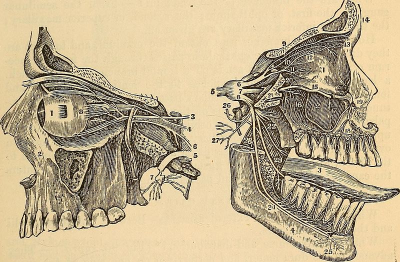
ANATOMY. 175 With what other nerves does it inosculate? — The ophthalmis andmaxillary. SECOND PAIR. Whence do the optic nerves arise ?^ — They arise from the thalaminervorum. What is their course and distribution ? — They pass outward andunite before the sella turcica, separate and leave the cranium throughthe optic foramen, and pass to enter the globe of the eye. THIRD PAIR. Whence do the third pair arise ? ^ (Figs. 95, 96,97.)—The third pair,or motores oculorum, arise from the crura cerebri, just before the an-terior edge of the pons varolii. Fig. 96. Fig. 97.
Text Appearing After Image:
What is their course? — They perforate the dura mater behind theposterior clynoid processes, run along the upper part of the cavernoussinus, and pass out through the foramen lacerum superius. Enumerate the branches of the motores oculi or third pair.—-Each sends a branch to the rectus superior, and a ramus to the levatorpalpebrae superioris; then a branch to the rectus internus; a branchto the rectus inferior; the longest branch to the obiiquus inferior;and a branch to the lenticular ganglion. What is the distribution of the ciliary plexus ? — From the lenti- 176 ANATOMY. cular gahgllon proceed several filiments, forming the ciliary plexus;which surround the optic nerve, and run to the iris, which theysupply. FOFKTH PAIR . Whence do the pathetic! arise?^—Behind the nates.What is their course and distribution ? ^ — They pass on each sideto the edge of the tentorium, and along the upper part of the cavern-ous sinus, and pass out through the foramen lacerum orbitale inferius,an

Note About Images

Please note that these images are extracted from scanned page images that may have been digitally enhanced for readability - coloration and appearance of these illustrations may not perfectly resemble the original work.
Date
Source

https://www.flickr.com/photos/internetarchivebookimages/14594360687/

Author Internet Archive Book Images
Permission
(Reusing this file)
At the time of upload, the image license was automatically confirmed using the Flickr API. For more information see Flickr API detail.
Flickr tags
InfoField
  • bookid:manualofexaminat00ludl
  • bookyear:1867
  • bookdecade:1860
  • bookcentury:1800
  • bookauthor:Ludlow__J__L___John_Livingston___1819_1888
  • booksubject:Medicine
  • bookpublisher:Philadelphia___Henry_C__Lea
  • bookcontributor:Francis_A__Countway_Library_of_Medicine
  • booksponsor:Open_Knowledge_Commons_and_Harvard_Medical_School
  • bookleafnumber:180
  • bookcollection:medicalheritagelibrary
  • bookcollection:francisacountwaylibrary
  • bookcollection:americana
Flickr posted date
InfoField
30 July 2014


Licensing edit

This image was taken from Flickr's The Commons. The uploading organization may have various reasons for determining that no known copyright restrictions exist, such as:
  1. The copyright is in the public domain because it has expired;
  2. The copyright was injected into the public domain for other reasons, such as failure to adhere to required formalities or conditions;
  3. The institution owns the copyright but is not interested in exercising control; or
  4. The institution has legal rights sufficient to authorize others to use the work without restrictions.

More information can be found at https://flickr.com/commons/usage/.


Please add additional copyright tags to this image if more specific information about copyright status can be determined. See Commons:Licensing for more information.
This image was originally posted to Flickr by Internet Archive Book Images at https://flickr.com/photos/126377022@N07/14594360687. It was reviewed on 26 September 2015 by FlickreviewR and was confirmed to be licensed under the terms of the No known copyright restrictions.

26 September 2015

File history

Click on a date/time to view the file as it appeared at that time.

Date/TimeThumbnailDimensionsUserComment
current15:02, 26 September 2015Thumbnail for version as of 15:02, 26 September 20151,916 × 1,256 (507 KB) (talk | contribs)== {{int:filedesc}} == {{information |description={{en|1=<br> '''Identifier''': manualofexaminat00ludl ([https://commons.wikimedia.org/w/index.php?title=Special%3ASearch&profile=default&fulltext=Search&search=insource%3A%2Fmanualofexaminat00ludl%2F fin...

There are no pages that use this file.