File:A red blood cell in a capillary, pancreatic tissue - TEM.jpg

Original file (1,560 × 1,254 pixels, file size: 579 KB, MIME type: image/jpeg)
Captions
Captions
Summary
edit
| Description |
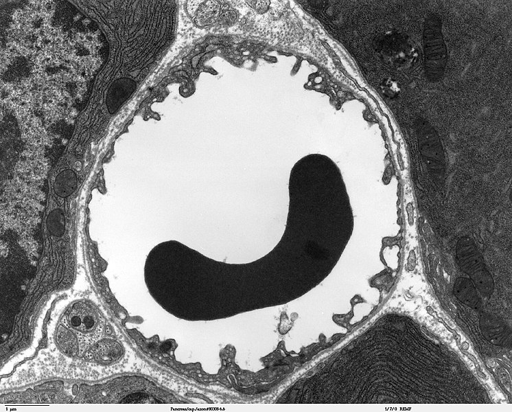
Transmission electron microscope image of a thin section cut through the pancreas(mammalian). This image shows a capillary within the pancreatic tissue(acinar cells in this image). Note the abundance of rough endoplasmic reticulum in the acinar cells. There is a red blood cell within the capillary. The capillary lining consists of long, thin endothelial cells, connected by tight junctions. The image shows fenestration of these endothelial cells. The image also shows synaptic vesicles in the neuron(nerve cell) next to the capillary. JEOL 100CX |
| Source | |
| Author | Louisa Howard |
| Permission (Reusing this file) |
PD |
Licensing
edit
| This work has been released into the public domain by its author, Louisa Howard. This applies worldwide. In some countries this may not be legally possible; if so: Louisa Howard grants anyone the right to use this work for any purpose, without any conditions, unless such conditions are required by law.
|
File history
Click on a date/time to view the file as it appeared at that time.
| Date/Time | Thumbnail | Dimensions | User | Comment | |
|---|---|---|---|---|---|
| current | 23:44, 4 October 2006 | | 1,560 × 1,254 (579 KB) | Patho (talk | contribs) | {{Information |Description=Transmission electron microscope image of a thin section cut through the pancreas(mammalian). This image shows a capillary within the pancreatic tissue(acinar cells in this image). Note the abundance of rough endoplasmic reticul |
You cannot overwrite this file.
File usage on Commons
There are no pages that use this file.
File usage on other wikis
The following other wikis use this file:
- Usage on bg.wikipedia.org
- Usage on bn.wikipedia.org
- Usage on bs.wikipedia.org
- Usage on de.wikipedia.org
- Usage on de.wikibooks.org
- Usage on en.wikipedia.org
- Usage on es.wikipedia.org
- Usage on eu.wikipedia.org
- Usage on fa.wikipedia.org
- Usage on fr.wikipedia.org
- Usage on gl.wikipedia.org
- Usage on it.wikipedia.org
- Usage on kk.wikipedia.org
- Usage on ko.wikipedia.org
- Usage on lv.wikipedia.org
- Usage on mk.wikipedia.org
- Usage on ml.wikipedia.org
- Usage on ms.wikipedia.org
- Usage on nl.wikipedia.org
- Usage on nn.wikipedia.org
- Usage on vi.wikipedia.org
- Usage on war.wikipedia.org
- Usage on www.wikidata.org
- Usage on zh.wikipedia.org