File:Anatomy of an IgG.png

Size of this preview: 546 × 599 pixels. Other resolutions: 219 × 240 pixels | 437 × 480 pixels | 700 × 768 pixels | 1,125 × 1,235 pixels.
Original file (1,125 × 1,235 pixels, file size: 194 KB, MIME type: image/png)
Captions
Captions
Add a one-line explanation of what this file represents
Summary edit
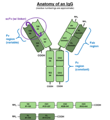
| Description | This image depicts the various domains and regions of a typical IgG. Note that residue addresses are approximate. |
| Source | Created by Wikipedia w:User:AJVincelli using PowerPoint 2013 and multiple public reference sources. |
| Author | w:User:AJVincelli |
Licensing edit
| This work has been released into the public domain by its author, AJVincelli at English Wikipedia. This applies worldwide. In some countries this may not be legally possible; if so: AJVincelli grants anyone the right to use this work for any purpose, without any conditions, unless such conditions are required by law. |
Original upload log edit
The original description page was here. All following user names refer to en.wikipedia.
| Date/Time | Dimensions | User | Comment |
|---|---|---|---|
| 2015-09-18 16:29:12 | 1125× 1235× | AJVincelli | Corrected scFv depiction |
| 2015-05-21 17:29:53 | 1125× 1235× | AJVincelli |
File history
Click on a date/time to view the file as it appeared at that time.
| Date/Time | Thumbnail | Dimensions | User | Comment | |
|---|---|---|---|---|---|
| current | 08:43, 27 December 2016 | | 1,125 × 1,235 (194 KB) | FastilyClone (talk | contribs) | Transferred from en.wikipedia (MTC!) |
You cannot overwrite this file.
File usage on Commons
There are no pages that use this file.
File usage on other wikis
The following other wikis use this file:
- Usage on bs.wikipedia.org
- Usage on en.wikipedia.org
- Usage on eu.wikipedia.org
- Usage on gl.wikipedia.org
- Usage on ja.wikipedia.org
- Usage on la.wikipedia.org
- Usage on ru.wikipedia.org
- Usage on th.wikipedia.org
- Usage on uz.wikipedia.org
- Usage on zh.wikipedia.org