File:Anatosuchus skull diagrams.jpg

Original file (1,512 × 1,599 pixels, file size: 1.02 MB, MIME type: image/jpeg)
Captions
Captions
Summary
edit
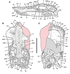
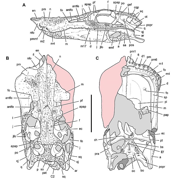
| Description |
Skull of the crocodyliform Anatosuchus minor. Drawings matching the skull (MNN GAD17) in Fig. 5. A Lateral view. B Dorsal view. C Ventral view. Pink tone indicates restored snout margin; parallel lines indicate broken bone surface; dashed line indicates missing bone; grey tone indicates matrix. Scale bar equals 5 cm. Abbreviations: a, angular; antfe, antorbital fenestra; antfo, antorbital fossa; apap, articular surface for palpebral; ar, articular; bo, basioccipital; bs, basisphenoid; C2, cervical vertebra 2 (axis); ch, choana; cqp, cranioquadrate passage; d, dentary; d1, dentary tooth 1; ec, ectopterygoid; Ef, Eustachian foramen; emf, external mandibular fenestra; en, external naris; f, frontal; fl , fl ange; fo, foramen; gef, groove for ear fl ap; j, jugal; jfo, jugal fossa; l, lacrimal; m, maxilla; m1, 2, 4, 17, maxillary tooth 1, 2, 4, 17; n, nasal; nfo, narial fossa; oc, occipital condyle; ot, otoccipital; p, parietal; pap, palpebral; pat, proatlas; pf, prefrontal; pl, palatine; pm, premaxilla; pm1, 6, premaxillary tooth 1, 6; pmmf, premaxilla-maxilla foramen; po, postorbital; popr, paroccipital process; pos, preotic siphonium; pra, prearticular; pt, pterygoid; q, quadrate; qj, quadratojugal; sa, surangular; sp, splenial; sq, squamosal; so, supraoccipital. |
|||
| Date | ||||
| Source | Cretaceous Crocodyliforms from the Sahara. ZooKeys 28: 1–143. doi:10.3897/zookeys.28.325 | |||
| Author | Sereno PC, Larsson HCE | |||
| Permission (Reusing this file) |
|
File history
Click on a date/time to view the file as it appeared at that time.
| Date/Time | Thumbnail | Dimensions | User | Comment | |
|---|---|---|---|---|---|
| current | 01:44, 11 August 2022 | | 1,512 × 1,599 (1.02 MB) | FunkMonk (talk | contribs) | == {{int:filedesc}} == {{Information |Description=Skull of the crocodyliform Anatosuchus minor. Drawings matching the skull (MNN GAD17) in Fig. 5. A Lateral view. B Dorsal view. C Ventral view. Pink tone indicates restored snout margin; parallel lines indicate broken bone surface; dashed line indicates missing bone; grey tone indicates matrix. Scale bar equals 5 cm. Abbreviations: a, angular; antfe, antorbital fenestra; antfo, antorbital fossa; apap, articular surface for palpebral; ar, artic... |
You cannot overwrite this file.
File usage on Commons
There are no pages that use this file.
File usage on other wikis
The following other wikis use this file:
- Usage on en.wikipedia.org