File:Apical cap Case 136 (4800110895).jpg

Size of this preview: 800 × 600 pixels. Other resolutions: 320 × 240 pixels | 640 × 480 pixels | 1,024 × 768 pixels | 1,280 × 960 pixels | 1,600 × 1,200 pixels.
Original file (1,600 × 1,200 pixels, file size: 814 KB, MIME type: image/jpeg)
Captions
Captions
Add a one-line explanation of what this file represents
Summary
edit.jpg&action=edit§ion=1)
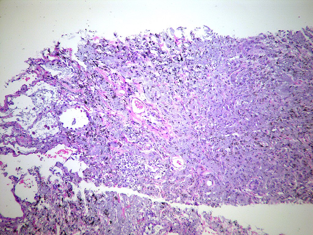
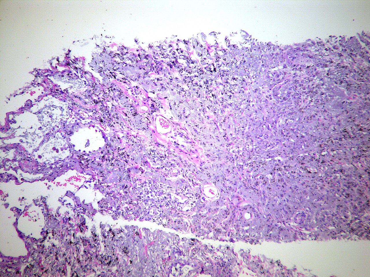
| Description | This low magnification view shows some alveoli at the left side of this apical cap. The apical cap is a pulmonary parenchymal lesion that often is accompanied by overlying pleural fibrosis. Needle core biopsy. Hematoxylin and eosin stain. |
| Date | |
| Source | Apical cap Case 136 |
| Author | Yale Rosen from USA |
Licensing
edit.jpg&action=edit§ion=2)
This file is licensed under the Creative Commons Attribution-Share Alike 2.0 Generic license.
- You are free:
- to share – to copy, distribute and transmit the work
- to remix – to adapt the work
- Under the following conditions:
- attribution – You must give appropriate credit, provide a link to the license, and indicate if changes were made. You may do so in any reasonable manner, but not in any way that suggests the licensor endorses you or your use.
- share alike – If you remix, transform, or build upon the material, you must distribute your contributions under the same or compatible license as the original.
| This image was originally posted to Flickr by Pulmonary Pathology at https://flickr.com/photos/30950973@N03/4800110895 (archive). It was reviewed on 3 October 2019 by FlickreviewR 2 and was confirmed to be licensed under the terms of the cc-by-sa-2.0. |
Category:Files_from_Yale_Rosen_Flickr_stream
File history
Click on a date/time to view the file as it appeared at that time.
| Date/Time | Thumbnail | Dimensions | User | Comment | |
|---|---|---|---|---|---|
| current | 10:14, 3 October 2019 | | 1,600 × 1,200 (814 KB) | Netha Hussain (talk | contribs) | Transferred from Flickr via #flickr2commons |
You cannot overwrite this file.
File usage on Commons
There are no pages that use this file.
.jpg&oldid=894603026)