File:Aristosuchus.jpg

Original file (2,190 × 3,048 pixels, file size: 2.81 MB, MIME type: image/jpeg)
Captions
Captions
Summary
edit
| Description |
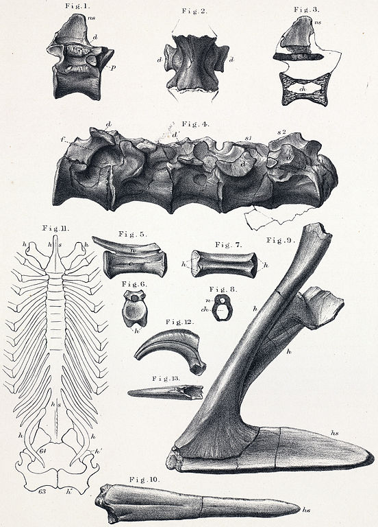
Poikilopleuron pusillus. Fig. 1. Side view of a dorsal vertebra. Fig. 2. Under view of a dorsal vertebra. Fig. 3. Vertical longitudinal section of the same. Fig. 4. Side view of lumbar and sacral vertebræ. Fig. 5. Side view of a caudal vertebra. Fig. 6. End view of the same. Fig. 7. Under view of the same. Fig. 8. Vertical transverse section of the middle of the same. Fig. 9. Abdominal hæmapophysis and hæmal spine. Fig. 10. Under surface of abdominal hæmal spine. Fig. 11. Reduced view, in outline, of neural or upper surface of the series of abdominal hæmapophyses and spines of a Crocodilus biporcatus. Fig. 12. Side view of ungual phalanx. Fig. 13. Upper view of the same. The fossils are of the natural size, are from the Wealden of the Isle of Wight, and are in the Museum of the Rev. W. Fox, M.A., F.G.S. |
| Date | 1800s |
| Source | A History of British Fossil Reptiles, Vol. II. by Sir Richard Owen. |
| Author | C. L. Griesbach |
Licensing
edit
|
This work is in the public domain in its country of origin and other countries and areas where the copyright term is the author's life plus 70 years or fewer.
| |
| This file has been identified as being free of known restrictions under copyright law, including all related and neighboring rights. | |
File history
Click on a date/time to view the file as it appeared at that time.
| Date/Time | Thumbnail | Dimensions | User | Comment | |
|---|---|---|---|---|---|
| current | 16:44, 30 March 2011 | | 2,190 × 3,048 (2.81 MB) | FunkMonk (talk | contribs) | {{Information |Description=Poikilopleuron pusillus. Fig. 1. Side view of a dorsal vertebra. Fig. 2. Under view of a dorsal vertebra. Fig. 3. Vertical longitudinal section of the same. Fig. 4. Side view of lumbar and sacral vertebræ. Fig. 5. Side vie |
You cannot overwrite this file.
File usage on Commons
The following page uses this file:
File usage on other wikis
The following other wikis use this file:
- Usage on en.wikipedia.org
- Usage on es.wikipedia.org
- Usage on sk.wikipedia.org
- Usage on sv.wikipedia.org
- Usage on www.wikidata.org
- Usage on zh.wikipedia.org