File:Basioccipital of Acamptonectes.png
Size of this preview: 644 × 600 pixels. Other resolutions: 258 × 240 pixels | 515 × 480 pixels | 825 × 768 pixels | 1,099 × 1,024 pixels | 2,028 × 1,889 pixels.
Original file (2,028 × 1,889 pixels, file size: 4.08 MB, MIME type: image/png)
Captions
Captions
Add a one-line explanation of what this file represents
Summary
edit
| Description |
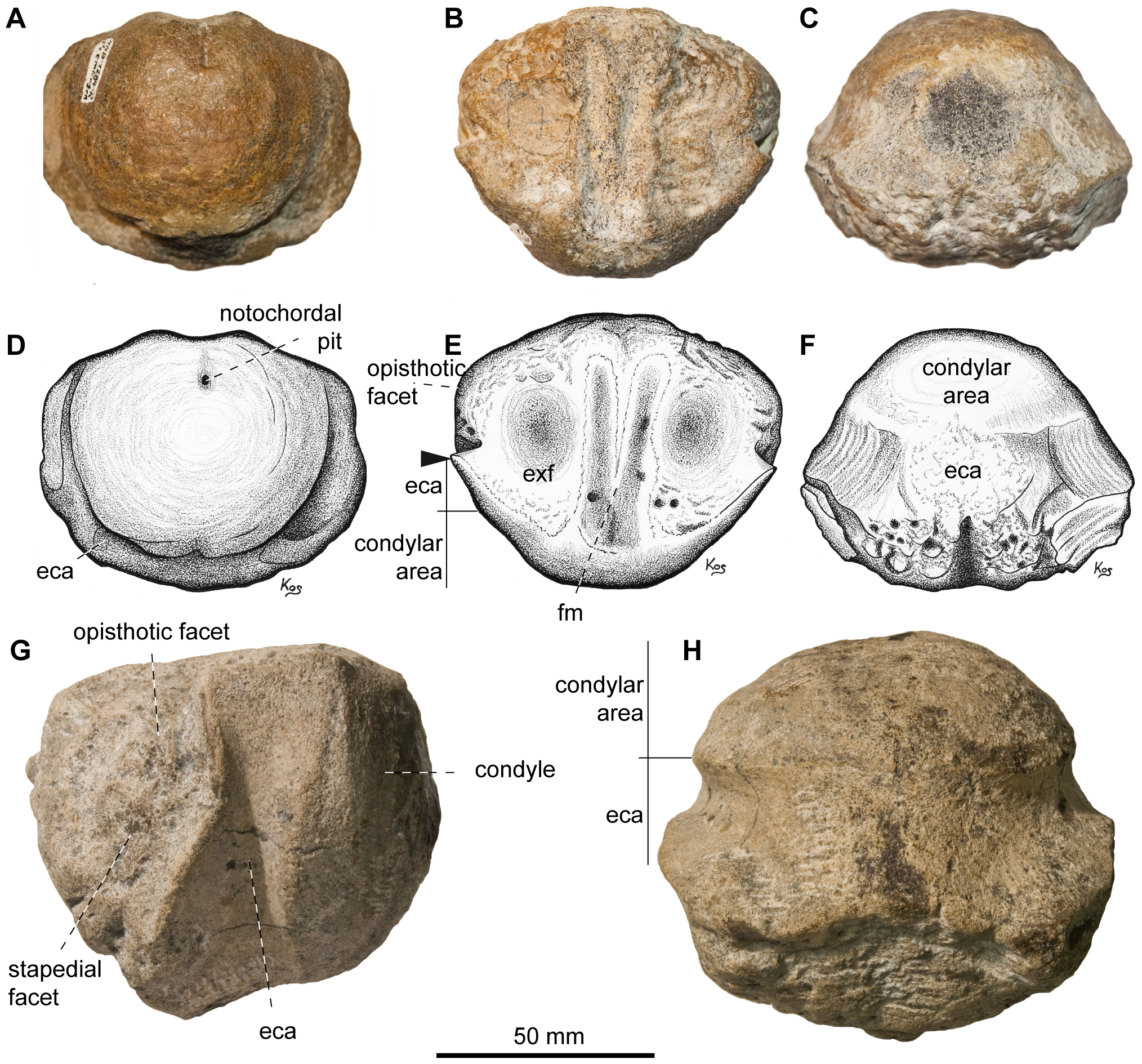
Basioccipital of Acamptonectes densus. A–F: SNHM1284-R, in posterior view (A,D), in dorsal view (B,E), and in ventral view (C,F). G–H: GLAHM132588 (holotype), in lateral view (G) and ventral view (H). Note the markedly concave extracondylar band that separates the condyle from the rest of the basioccipital and the bilobed median concavity for the foramen magnum. The arrow in E indicates the protruding anterior edge of the extracondylar area posterior to the depressed opisthotic facet. Abbreviations: eca: extracondylar area; exf: exoccipital facet; fm: median concavity for the foramen magnum. |
||
| Date | |||
| Source | http://www.plosone.org/article/info%3Adoi%2F10.1371%2Fjournal.pone.0029234 | ||
| Author | Valentin Fischer, Michael W. Maisch, Darren Naish, Ralf Kosma, Jeff Liston, Ulrich Joger, Fritz J. Krüger, Judith Pardo Pérez, Jessica Tainsh, Robert M. Appleby† | ||
| Permission (Reusing this file) |
|
File history
Click on a date/time to view the file as it appeared at that time.
| Date/Time | Thumbnail | Dimensions | User | Comment | |
|---|---|---|---|---|---|
| current | 21:15, 18 July 2019 | | 2,028 × 1,889 (4.08 MB) | FunkMonk (talk | contribs) | == {{int:filedesc}} == {{Information |Description=Basioccipital of Acamptonectes densus. A–F: SNHM1284-R, in posterior view (A,D), in dorsal view (B,E), and in ventral view (C,F). G–H: GLAHM132588 (holotype), in lateral view (G) and ventral view (H). Note the markedly concave extracondylar band that separates the condyle from the rest of the basioccipital and the bilobed median concavity for the foramen magnum. The arrow in E indicates the protruding anterior edge of the extracondylar area p... |
You cannot overwrite this file.
File usage on Commons
There are no pages that use this file.
File usage on other wikis
The following other wikis use this file:
- Usage on en.wikipedia.org
- Usage on nl.wikipedia.org