File:Blood clot in scanning electron microscopy.jpg
No higher resolution available.
Blood_clot_in_scanning_electron_microscopy.jpg (700 × 475 pixels, file size: 76 KB, MIME type: image/jpeg)
Captions
Captions
Add a one-line explanation of what this file represents
Summary
edit
| Description |
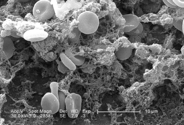
English: This scanning electron micrograph (SEM) depicted a number of red blood cells found enmeshed in a fibrinous matrix on the luminal surface of an indwelling vascular catheter; Magnified 2858x.
Note the biconcave cytomorphologic shape of each erythrocyte, which increases the surface area of these hemoglobin-filled cells, thereby, promoting a greater degree of gas exchange, which is their primary function in an in vivo setting. In their adult phase, these cells possess no nucleus. What appears to be irregularly-shaped chunks of debris, are actually fibrin clumps, which when inside the living organism, functions as a key component in the process of blood clot formation, acting to entrap the red blood cells in a mesh-like latticework of proteinaceous strands, thereby, stabilizing and strengthening the clot, in much the same way as rebar acts to strengthen, and reinforce cement. |
| Date | |
| Source | http://phil.cdc.gov/phil/details.asp |
| Author | Janice Carr |
Licensing
edit
| This work has been released into the public domain by its author, http://phil.cdc.gov/phil/details.asp Janice Carr. This applies worldwide. In some countries this may not be legally possible; if so: http://phil.cdc.gov/phil/details.asp Janice Carr grants anyone the right to use this work for any purpose, without any conditions, unless such conditions are required by law.
|
File history
Click on a date/time to view the file as it appeared at that time.
| Date/Time | Thumbnail | Dimensions | User | Comment | |
|---|---|---|---|---|---|
| current | 17:25, 13 May 2015 | | 700 × 475 (76 KB) | Jean-madeleine de sainte agathe (talk | contribs) | User created page with UploadWizard |
You cannot overwrite this file.
File usage on Commons
There are no pages that use this file.
File usage on other wikis
The following other wikis use this file:
- Usage on fr.wikipedia.org
- Usage on zh-yue.wikipedia.org
- Usage on zh.wikipedia.org