File:Bronchiolar area cilia cross-sections 1.jpg

Size of this preview: 751 × 600 pixels. Other resolutions: 301 × 240 pixels | 601 × 480 pixels | 962 × 768 pixels | 1,280 × 1,022 pixels | 1,600 × 1,278 pixels.
Original file (1,600 × 1,278 pixels, file size: 776 KB, MIME type: image/jpeg)
Captions
Captions
Add a one-line explanation of what this file represents
Summary
edit
| Description |
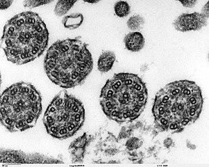
Transmission electron microscope image of a thin section cut through the bronchiolar area of lung(mouse), showing cilia cross-sections. The inside of the cilium contain precisely arranged microtubles, the axoneme. The axoneme has a central unit containing two single microtubules and nine peripheral doublet microtubules (known as the "9+2"). Dynein sidearms project from the A tubule of each doublet. JEOL 100CX TEM |
| Source | |
| Author | Louisa Howard, Michael Binder |
| Permission (Reusing this file) |
PD |
Licensing
edit
| This work has been released into the public domain by its author, Louisa Howard and Michael Binder. This applies worldwide. In some countries this may not be legally possible; if so: Louisa Howard and Michael Binder grants anyone the right to use this work for any purpose, without any conditions, unless such conditions are required by law.
|
File history
Click on a date/time to view the file as it appeared at that time.
| Date/Time | Thumbnail | Dimensions | User | Comment | |
|---|---|---|---|---|---|
| current | 14:38, 7 October 2006 | | 1,600 × 1,278 (776 KB) | Patho (talk | contribs) | {{Information |Description=Transmission electron microscope image of a thin section cut through the bronchiolar area of lung(mouse), showing cilia cross-sections. The inside of the cilium contain precisely arranged microtubles, the axoneme. The axoneme ha |
You cannot overwrite this file.
File usage on Commons
There are no pages that use this file.
File usage on other wikis
The following other wikis use this file:
- Usage on de.wikibooks.org