File:Cajal Retina.jpg
Original file (500 × 745 pixels, file size: 83 KB, MIME type: image/jpeg)
Captions
Captions
Summary edit
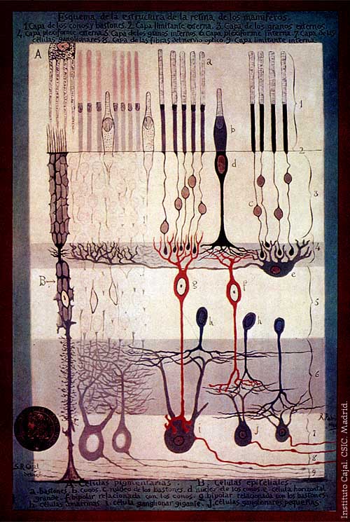
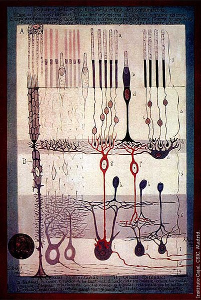
From "Structure of the Mammalian Retina" c.1900 By Santiago Ramon y Cajal.
Outline of the structure of the mammalian retina. 1. Rod and cone layer. 2. External limiting membrane. 3. Outer granular layer. 4. Outer plexiform layer. 5. Inner granular layer. 6. Inner plexiform layer. 7. Ganglion cell layer. 8. Optic nerve fibre layer. 9. Internal limiting membrane. A. Pigmented cells. B. Epithelial cells. a. Rods. b. Cones. c. Rod nucleus. d. Cone Nucleus. e. Large horizontal cell f. Cone-associated bipolar cell. g. Rod-associated bipolar cell. h. Amacrine cells. i. Giant ganglion cell. j. Small ganglion cells.
Licensing edit
|
This work is in the public domain in its country of origin and other countries and areas where the copyright term is the author's life plus 70 years or fewer.
| |
| This file has been identified as being free of known restrictions under copyright law, including all related and neighboring rights. | |
File history
Click on a date/time to view the file as it appeared at that time.
| Date/Time | Thumbnail | Dimensions | User | Comment | |
|---|---|---|---|---|---|
| current | 17:42, 4 March 2006 | | 500 × 745 (83 KB) | Feezil~commonswiki (talk | contribs) | From "Structure of the Mammalian Retina" c.1900 By Santiago Ramon y Cajal. 1.- Rod and Cone layer 2.-Outer nuclear layer 3.- Granule layer 4.- External plexiform layer A: Pigmented cells; B: epithelial cells |
You cannot overwrite this file.
File usage on Commons
There are no pages that use this file.
File usage on other wikis
The following other wikis use this file:
- Usage on ar.wikipedia.org
- Usage on bn.wikipedia.org
- Usage on ca.wikipedia.org
- Usage on en.wikipedia.org
- Usage on en.wikiversity.org
- Human vision and function/Part 1: How the eye works/1.3 Light stimulus and the eye
- User:Jtwsaddress42/People/Ramón y Cajal, Santiago
- User:Jtwsaddress42/People/R
- User:Jtwsaddress42/Gallery/Ramón y Cajal, Santiago
- User:Jtwsaddress42/Gallery/Ramón y Cajal, Santiago - The Visual System
- User:Jtwsaddress42/Gallery
- Usage on es.wikipedia.org
- Usage on et.wikipedia.org
- Usage on ext.wikipedia.org
- Usage on fa.wikipedia.org
- Usage on fr.wikipedia.org
- Usage on gl.wikipedia.org
- Usage on he.wikipedia.org
- Usage on hy.wikipedia.org
- Usage on it.wikipedia.org
- Usage on ja.wikipedia.org
- Usage on ko.wikipedia.org
- Usage on ml.wikipedia.org
- Usage on pt.wikipedia.org
- Usage on ru.wikipedia.org
- Usage on simple.wikipedia.org
- Usage on th.wikipedia.org
- Usage on uk.wikipedia.org
- Usage on vi.wikipedia.org
- Usage on zh.wikipedia.org