File:Chlamydomanas reinhardtii Flagella 5 - TEM.jpg

Original file (1,800 × 1,438 pixels, file size: 650 KB, MIME type: image/jpeg)
Captions
Captions
Summary
edit
| Description |
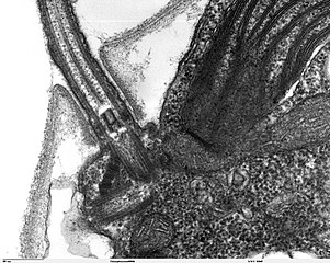
Transmission electron microscope image, showing an example of green algae (Chlorophyta). Chlamydomanas reinhardtii is a unicellular flagellate used as a model system in molecular genetics work and flagellar motility studies. This image is a longitudinal section through a portion of the flagellar apparatus. In the cell apex are the basal body regions that are the anchoring sites for the flagella. This image shows that the two flagella form a V and they are connected at their bases by a transversely striated fibre. This connection is thought to play a part in the coordination of flagellar movement. Also visible is the transition region, with its fibers of the stellate structure. JEOL 100CX TEM |
| Source | |
| Author | Elizabeth Smith, Louisa Howard, Erin Dymek |
| Permission (Reusing this file) |
PD |
Licensing
edit
| This work has been released into the public domain by its author, Elizabeth Smith, Louisa Howard and Erin Dymek. This applies worldwide. In some countries this may not be legally possible; if so: Elizabeth Smith, Louisa Howard and Erin Dymek grants anyone the right to use this work for any purpose, without any conditions, unless such conditions are required by law.
|
File history
Click on a date/time to view the file as it appeared at that time.
| Date/Time | Thumbnail | Dimensions | User | Comment | |
|---|---|---|---|---|---|
| current | 14:01, 7 October 2006 | | 1,800 × 1,438 (650 KB) | Patho (talk | contribs) | {{Information |Description=Transmission electron microscope image, showing an example of green algae (Chlorophyta). Chlamydomanas reinhardtii is a unicellular flagellate used as a model system in molecular genetics work and flagellar motility studies. T |
You cannot overwrite this file.
File usage on Commons
There are no pages that use this file.
File usage on other wikis
The following other wikis use this file:
- Usage on de.wikibooks.org
- Usage on ja.wikipedia.org
- Usage on uk.wikipedia.org