File:Chlamydomonas6-1.jpg
Size of this preview: 585 × 599 pixels. Other resolutions: 234 × 240 pixels | 469 × 480 pixels | 750 × 768 pixels | 1,024 × 1,049 pixels.
Original file (1,024 × 1,049 pixels, file size: 386 KB, MIME type: image/jpeg)
Captions
Captions
Add a one-line explanation of what this file represents
| Description |
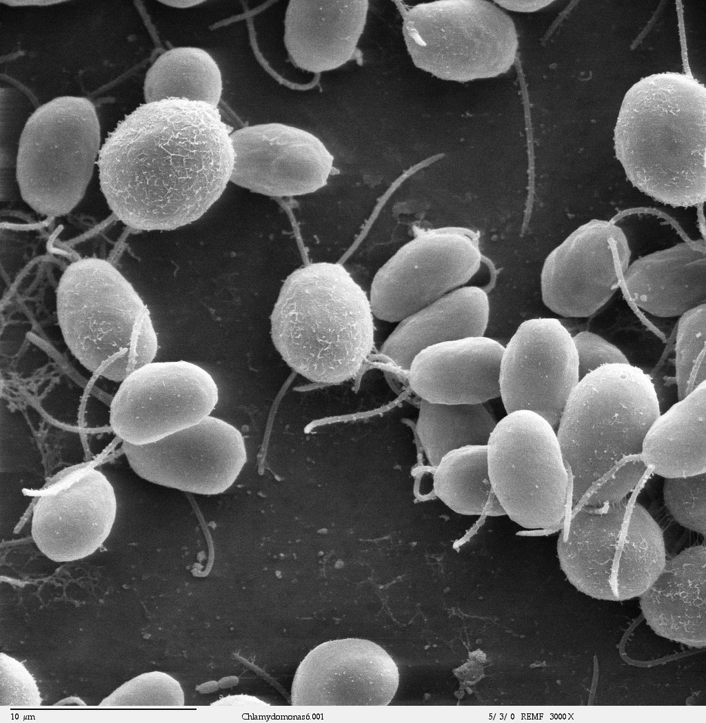
Scanning electron microscope image, showing an example of green algae (Chlorophyta). Chlamydomanas reinhardtii is a unicellular flagellate used as a model system in molecular genetics work and flagellar motility studies. Smith, E.F and P.A. Lefebvre (1996) "PF16 Encodes a Protein with Armadillo Repeats and Localizes to a Single Microtubule of the Central Apparatus in Chlamydomonas Flagella", J. Cell Biology, 132(3): 359-370 |
| Date | |
| Source | Source and public domain notice at http://remf.dartmouth.edu/imagesindex.html |
| Author | Dartmouth Electron Microscope Facility, Dartmouth College |
| Permission (Reusing this file) |
Released into the public domain |
| This work has been released into the public domain by its author, Dartmouth Electron Microscope Facility, Dartmouth College. This applies worldwide. In some countries this may not be legally possible; if so: Dartmouth Electron Microscope Facility, Dartmouth College grants anyone the right to use this work for any purpose, without any conditions, unless such conditions are required by law.
|
File history
Click on a date/time to view the file as it appeared at that time.
| Date/Time | Thumbnail | Dimensions | User | Comment | |
|---|---|---|---|---|---|
| current | 06:19, 21 September 2007 | | 1,024 × 1,049 (386 KB) | Neil916 (talk | contribs) | {{Information |Description=en:Chlamydomonas, a single-celled green en:alga. |Source=Source and public domain notice at http://remf.dartmouth.edu/imagesindex.html |Date=20 September, 2007 |Author=Dartmouth Electron Microscope Facility, Dartmouth Co |
You cannot overwrite this file.
File usage on Commons
The following 2 pages use this file:
File usage on other wikis
The following other wikis use this file:
- Usage on ar.wikipedia.org
- Usage on arz.wikipedia.org
- Usage on ca.wikipedia.org
- Usage on ceb.wikipedia.org
- Usage on cs.wikipedia.org
- Usage on de.wikipedia.org
- Usage on el.wikipedia.org
- Usage on en.wikipedia.org
- Usage on en.wiktionary.org
- Usage on eo.wikipedia.org
- Usage on es.wikipedia.org
- Usage on es.wiktionary.org
- Usage on eu.wikipedia.org
- Usage on gl.wikipedia.org
- Usage on hu.wikipedia.org
- Usage on hu.wikibooks.org
- Usage on hy.wikipedia.org
- Usage on id.wikipedia.org
- Usage on it.wikipedia.org
- Usage on ko.wikipedia.org
- Usage on la.wikipedia.org
- Usage on nn.wikipedia.org
- Usage on pl.wikipedia.org
- Usage on pt.wikipedia.org
- Usage on ru.wikipedia.org
- Usage on sk.wikipedia.org
- Usage on species.wikimedia.org
- Usage on tr.wikipedia.org
- Usage on uk.wikipedia.org
- Usage on vi.wikipedia.org
View more global usage of this file.