File:Chromopsin Phylogeny with the Peropsins Highlighted.svg

Size of this PNG preview of this SVG file: 800 × 497 pixels. Other resolutions: 320 × 199 pixels | 640 × 398 pixels | 1,024 × 637 pixels | 1,280 × 796 pixels | 2,560 × 1,592 pixels | 2,300 × 1,430 pixels.
Original file (SVG file, nominally 2,300 × 1,430 pixels, file size: 129 KB)
Captions
Captions
Chromopsin Phylogeny with the Peropsins Highlighted
Summary
edit
| Description |
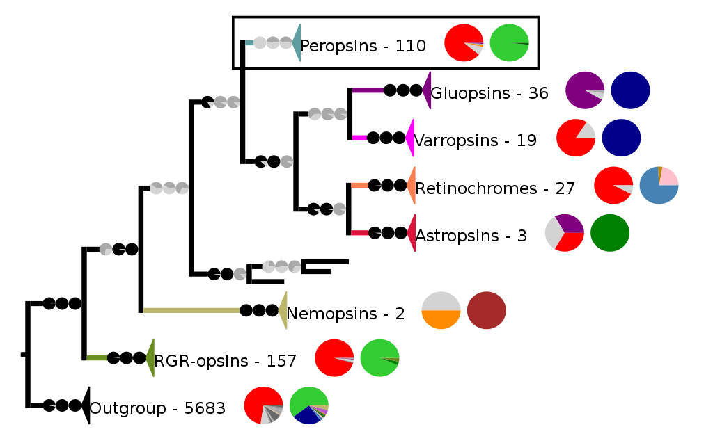
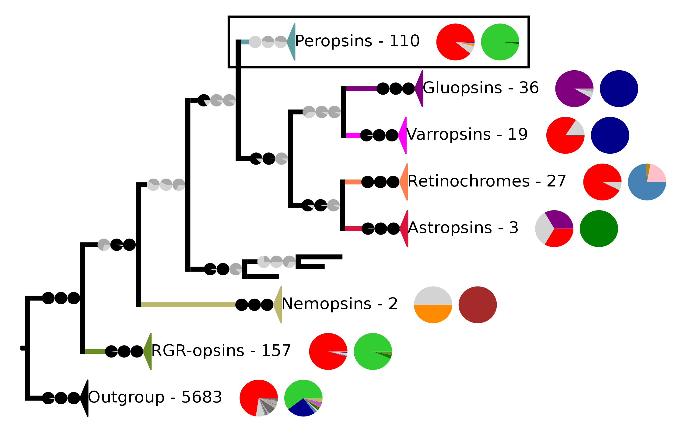
English: Phylogenetic reconstruction of the chromopsins. The main clades and the outgroup are collapsed. The outgroup contains non-opsin GPCRs and all other opsins. The frame highlights the peropsins. Next to each clade is shown the number of sequences within that clade. The first pie chart shows the percentage of a certain amino acid at lysine 2967.43. Red stands for lysine (K), purple stands for glutamic acid (E), orange stands for argenine (R), the other amino acids are alternatively colored dark or mid-gray so that two adjacent amino acids have different shades of gray in the pie chart. Light gray stands for a gap at this position as not all sequences have data there. The second pie chart gives the taxon composition for each clade, light green stands for craniates, dark green for cephalochordates, brown for nematodes, pale pink for annelids, dark blue for arthropods, and light blue for mollusks.
The support values are given as pie charts. They are from right to left SH-aLRT/aBayes/UFBoot. Splits are considered supported when SH-aLRT ≥ 80%, aBayes ≥ 0.95, and UFBoot ≥ 95%. If a support value is above its threshold the pie chart is black otherwise gray. |
| Date | |
| Source | SVG version of figure 2c with the peropsins highlighted instead of the gluopsins from the article: The Gluopsins: Opsins without the Retinal Binding Lysine, Cells 2022, 11(15), 2441; https://doi.org/10.3390/cells11152441 |
| Author | Martin Gühmann, Megan L. Porter, Michael J. Bok |
Licensing
edit
This file is licensed under the Creative Commons Attribution 4.0 International license.
- You are free:
- to share – to copy, distribute and transmit the work
- to remix – to adapt the work
- Under the following conditions:
- attribution – You must give appropriate credit, provide a link to the license, and indicate if changes were made. You may do so in any reasonable manner, but not in any way that suggests the licensor endorses you or your use.
File history
Click on a date/time to view the file as it appeared at that time.
| Date/Time | Thumbnail | Dimensions | User | Comment | |
|---|---|---|---|---|---|
| current | 12:30, 17 August 2022 | | 2,300 × 1,430 (129 KB) | Martin Gühmann (talk | contribs) | Uploaded a work by Martin Gühmann, Megan L. Porter, Michael J. Bok from SVG version of figure 2c from the article: The Gluopsins: Opsins without the Retinal Binding Lysine, Cells 2022, 11(15), 2441; https://doi.org/10.3390/cells11152441 with UploadWizard |
You cannot overwrite this file.
File usage on Commons
There are no pages that use this file.
File usage on other wikis
The following other wikis use this file:
- Usage on en.wikipedia.org