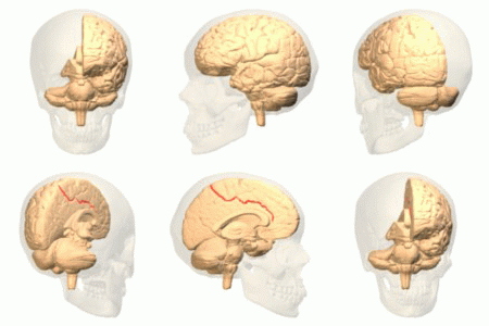
File:Cingulate sulcus animation.gif
Cingulate_sulcus_animation.gif (300 × 300 pixels, file size: 1.74 MB, MIME type: image/gif, looped, 72 frames, 7.2 s)
File information
Structured data
Captions
Captions
Add a one-line explanation of what this file represents
| DescriptionCingulate sulcus animation.gif |
English: Cingulate sulcus. One of sulci located in medial wall.
日本語: 左大脳半球の帯状溝。内側面に位置する脳溝のひとつ。 |
| Date | |
| Source | Polygon data are from BodyParts3D[1] |
| Author | Polygon data were generated by Database Center for Life Science(DBCLS)[2]. |
| Permission (Reusing this file) |
CC-BY-SA-2.1-jp |
| Other versions |
 |
This image was created with Blender.
This file is licensed under the Creative Commons Attribution-Share Alike 2.1 Japan license.
 |
  This image was made out of, or made from, content published in a BodyParts3D/Anatomography web site. The content of their website is published under the Creative Commons Attribution 2.1 Japan license. The author and licenser of the contents is
You can download 3D-polygon data of whole human body. And you can also manipulate and edit the polygon data using 3D softwares, for example, Meshlab or Blender. |
File history
Click on a date/time to view the file as it appeared at that time.
| Date/Time | Thumbnail | Dimensions | User | Comment | |
|---|---|---|---|---|---|
| current | 16:17, 21 February 2010 |  | 300 × 300 (1.74 MB) | Was a bee (talk | contribs) | {{Information |Description={{en|1= Cingulate sulcus. One of sulci located in medial wall. }} {{ja|1=左大脳半球の帯状溝。内側面に位置する脳溝のひとつ。 }} |Source=Polygon data are from BodyParts3D[http://lifesciencedb.jp/ag/bp3d/do |
You cannot overwrite this file.
File usage on Commons
The following page uses this file:
File usage on other wikis
The following other wikis use this file:
- Usage on ar.wikipedia.org
- Usage on pl.wikipedia.org
