File:DTI-sagittal-xyzrgb.jpg

Size of this preview: 544 × 599 pixels. Other resolutions: 218 × 240 pixels | 436 × 480 pixels | 1,021 × 1,125 pixels.
Original file (1,021 × 1,125 pixels, file size: 64 KB, MIME type: image/jpeg)
Captions
Captions
Add a one-line explanation of what this file represents
Summary
edit
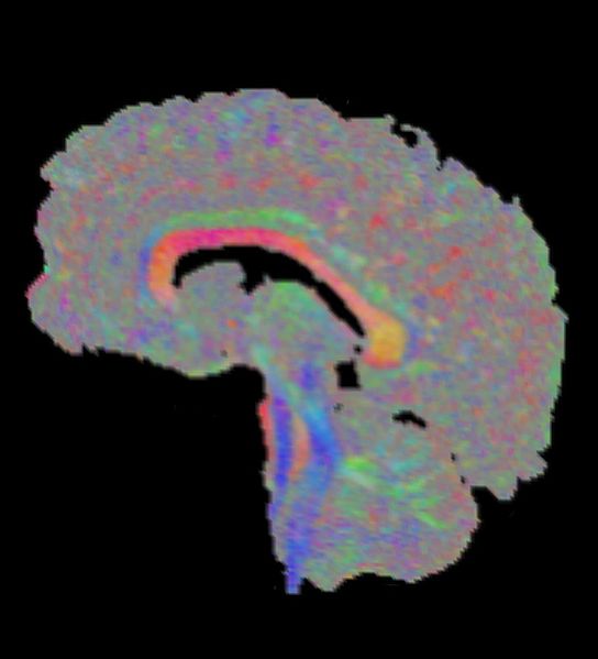
| Description | Visualization of DTI data, depicting a sagittal slice of a human brain near the mid-sagittal plane. The visualization uses the standard XYZ-RGB principal eigenvector color mapping. Especially prominent are the corpus callosum (large red structure in the center) and the fiber tracts that descend toward the spine (blue). Black areas are air or cavities that are filled with corticospinal fluid. |
| Date | |
| Source | Own work |
| Author | Thomas Schultz |
| Permission (Reusing this file) |
Rendering is own work, using a modified version of the BioTensor application developed at the University of Utah. The dataset is courtesy of Gordon Kindlmann at the Scientific Computing and Imaging Institute, University of Utah, and Andrew Alexander, W.M. Keck Laboratory for Functional Brain Imaging and Behaviour, University of Wisconsin, Madison. It is publicly available from [1] |
Licensing
edit
I, the copyright holder of this work, hereby publish it under the following licenses:

|
Permission is granted to copy, distribute and/or modify this document under the terms of the GNU Free Documentation License, Version 1.2 or any later version published by the Free Software Foundation; with no Invariant Sections, no Front-Cover Texts, and no Back-Cover Texts. A copy of the license is included in the section entitled GNU Free Documentation License. |
| This file is licensed under the Creative Commons Attribution-Share Alike 3.0 Unported license. | ||
| ||
| This licensing tag was added to this file as part of the GFDL licensing update. |
This file is licensed under the Creative Commons Attribution-Share Alike 2.5 Generic, 2.0 Generic and 1.0 Generic license.
- You are free:
- to share – to copy, distribute and transmit the work
- to remix – to adapt the work
- Under the following conditions:
- attribution – You must give appropriate credit, provide a link to the license, and indicate if changes were made. You may do so in any reasonable manner, but not in any way that suggests the licensor endorses you or your use.
- share alike – If you remix, transform, or build upon the material, you must distribute your contributions under the same or compatible license as the original.
You may select the license of your choice.
File history
Click on a date/time to view the file as it appeared at that time.
| Date/Time | Thumbnail | Dimensions | User | Comment | |
|---|---|---|---|---|---|
| current | 16:14, 22 September 2006 | | 1,021 × 1,125 (64 KB) | Thomas Schultz (talk | contribs) | {{Information |Description=Visualization of DTI data, depicting a sagittal slice of a human brain near the mid-sagittal plane. The visualization uses the standard XYZ-RGB principal eigenvector color mapping. Especially prominent are the corpus callosum (l |
You cannot overwrite this file.
File usage on Commons
The following page uses this file:
File usage on other wikis
The following other wikis use this file:
- Usage on de.wikipedia.org