File:Dorsal Root Galglia Neurons.tif
Size of this JPG preview of this TIF file: 800 × 250 pixels. Other resolutions: 320 × 100 pixels | 640 × 200 pixels | 1,024 × 320 pixels | 1,280 × 400 pixels | 6,144 × 1,920 pixels.
Original file (6,144 × 1,920 pixels, file size: 8.55 MB, MIME type: image/tiff)
Captions
Captions
Add a one-line explanation of what this file represents
Summary edit
| Description |
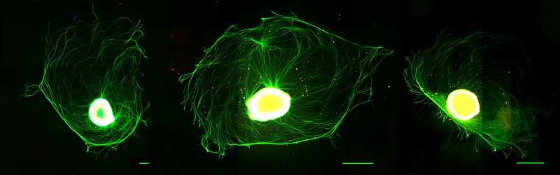
English: These cosmic thingies are the spinal ganglia (bulb with ~ 10,000 neurons) with radially growing axons. They were extracted from the spinal cord of a newborn rat. They grew on a special flat scaffold (polycaprolactone), were fed for 7 days, and finally sacrificed in the name of science (by using formaldehyde solution). Eventually, they underwent immunostaining using three fluorophores (DAPI, S100, anti-beta3-tubulin), and final scanning with imaging via fluorescence Olympus microscope.
This work was performed at the Centre for Cell Engineering, University of Glasgow, Scotland, UK.
Українська: Ось ці космічні штуковини - спінальні нервові вузли, або ганглії (центральна бульба з ~ 10 000 нейронами), з радіально виходячими з них аксонами, экстраговані зі спинного мозку новонароджених щурів (2-3 дні). Були посаджені на спеціальну плоску підкладку (полікапролактон), годовані протягом 7 днів, після чого були вбиті розчином формальдегіду в ім'я науки. Пройшли чотирьохденну стадію імунофарбування з використанням трьох флуорофорів (DAPI, S100, anti-beta3-tubulin), потім були сфотографовані за допомогою методу флуоресцентної мікроскопії (Olympus).
Робота була виконана в Центрі Клітинної Iнженерії Університету Глазго (Шотландія, Велика Британія). English: DRG Neurons |
| Date | |
| Source | Own work |
| Author | Kseniia Bondarenko |
Licensing edit
I, the copyright holder of this work, hereby publish it under the following license:
This file is licensed under the Creative Commons Attribution-Share Alike 4.0 International license.
- You are free:
- to share – to copy, distribute and transmit the work
- to remix – to adapt the work
- Under the following conditions:
- attribution – You must give appropriate credit, provide a link to the license, and indicate if changes were made. You may do so in any reasonable manner, but not in any way that suggests the licensor endorses you or your use.
- share alike – If you remix, transform, or build upon the material, you must distribute your contributions under the same or compatible license as the original.
|
This image was uploaded as part of Science Photo Competition 2016 |
File history
Click on a date/time to view the file as it appeared at that time.
| Date/Time | Thumbnail | Dimensions | User | Comment | |
|---|---|---|---|---|---|
| current | 20:09, 28 October 2016 | 6,144 × 1,920 (8.55 MB) | Morne Arin (talk | contribs) | User created page with UploadWizard |
You cannot overwrite this file.
File usage on Commons
The following page uses this file:
File usage on other wikis
The following other wikis use this file:
- Usage on ua.wikimedia.org
- Usage on uk.wikipedia.org