File:Ebstein8.jpg
Size of this preview: 800 × 334 pixels. Other resolutions: 320 × 134 pixels | 896 × 374 pixels.
Original file (896 × 374 pixels, file size: 51 KB, MIME type: image/jpeg)
Captions
Captions
Add a one-line explanation of what this file represents
Summary
edit
| Description |
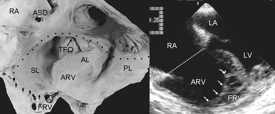
English: Internal view of right chambers. There is anatomic continuity between the septal and posterior leaflets tethered to the right ventricular wall, forming a cul-de-sac (arrows) with reduction of the apical portion of the ventricle, which is occupied by multiple trabeculae. The echocardiogram shows similar features to those seen in the anatomic specimen. TFO: Tricuspid functional opening. Other abbreviations as before. |
| Date | |
| Source | Muñoz-Castellanos L, Espinola-Zavaleta N, Kuri-Nivón M, Keirns C. Ebstein's Anomaly: anatomo-echocardiographic correlation. Cardiovasc Ultrasound. 5, 43. 2008. doi:10.1186/1476-7120-5-43. PMID 18034907. |
| Author | Luis Muñoz-Castellanos et al |
| Permission (Reusing this file) |
[1] |
Licensing
edit
This file is licensed under the Creative Commons Attribution 2.0 Generic license.
- You are free:
- to share – to copy, distribute and transmit the work
- to remix – to adapt the work
- Under the following conditions:
- attribution – You must give appropriate credit, provide a link to the license, and indicate if changes were made. You may do so in any reasonable manner, but not in any way that suggests the licensor endorses you or your use.
File history
Click on a date/time to view the file as it appeared at that time.
| Date/Time | Thumbnail | Dimensions | User | Comment | |
|---|---|---|---|---|---|
| current | 19:00, 25 June 2008 | | 896 × 374 (51 KB) | Filip em (talk | contribs) | == Opis == {{Information |Description={{en|1=Internal view of right chambers. There is anatomic continuity between the septal and posterior leaflets tethered to the right ventricular wall, forming a cul-de-sac (arrows) with reduction of the apical portion |
You cannot overwrite this file.
File usage on Commons
There are no pages that use this file.