File:Fiona pinnata 4.png
Size of this preview: 799 × 363 pixels. Other resolutions: 320 × 145 pixels | 640 × 291 pixels | 1,024 × 465 pixels | 1,280 × 582 pixels | 2,553 × 1,160 pixels.
Original file (2,553 × 1,160 pixels, file size: 1.28 MB, MIME type: image/png)
Captions
Captions
Add a one-line explanation of what this file represents
| Description |
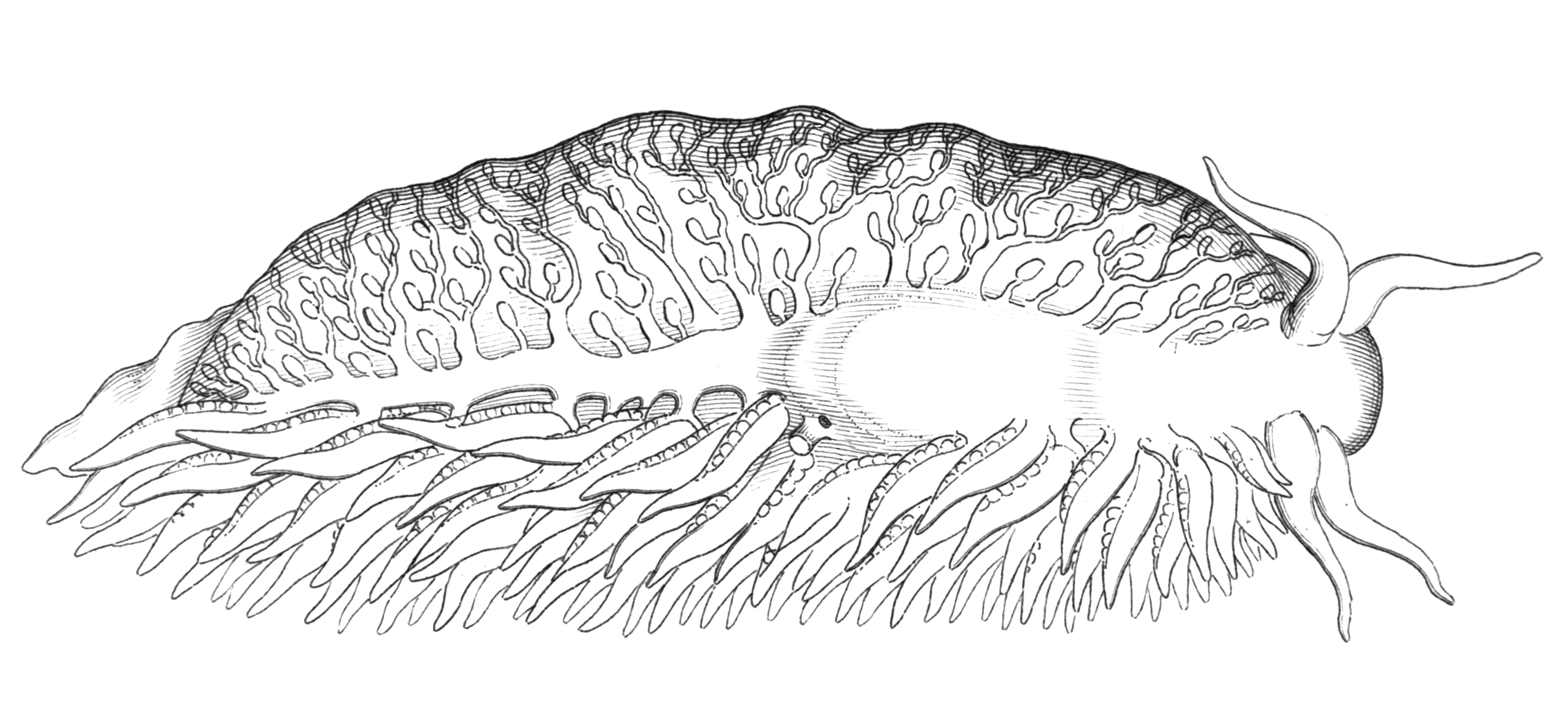
English: Drawing of dorsal view of the Fiona pinnata with cerata removed on the left side. There visible efferent vessels leading from cerata to branchio-cardiac vessel. There is also visible heart in the centre. Anus is visible as tube like opening next to the heart. There is also other opening next to anus, which probably leads into the kidney. |
| Date | |
| Source | Alder J. & Hancock A. (October 1851). "Descriptions of two new species of nudibranchiate Mollusca, one of them forming the type of a new genus". Annals and Magazine of Natural History (series 2)8(46): 290-302, pls. 9-10. Cropped from Plate 9 figure 2 and retouched by User:Snek01. |
| Author | Joshua Alder (1792-1867) & Albany Hancock (1806-1873) |
|
This work is in the public domain in its country of origin and other countries and areas where the copyright term is the author's life plus 100 years or fewer. | |
| This file has been identified as being free of known restrictions under copyright law, including all related and neighboring rights. | |
| Annotations | This image is annotated: View the annotations at Commons |
File history
Click on a date/time to view the file as it appeared at that time.
| Date/Time | Thumbnail | Dimensions | User | Comment | |
|---|---|---|---|---|---|
| current | 19:06, 18 December 2009 | | 2,553 × 1,160 (1.28 MB) | Snek01 (talk | contribs) | {{Information |Description={{en|Drawing of dorsal view of the ''Fiona pinnata'' with cerata removed on the left side. There visible efferent vessels leading from cerata to branchio-cardiac vessel. There is also visible heart in the centre. Anus is visible |
You cannot overwrite this file.
File usage on Commons
There are no pages that use this file.
File usage on other wikis
The following other wikis use this file:
- Usage on en.wikipedia.org