File:FocusStack BrightFieldLightMicroscopy DiatomaceousEarth.jpg

Size of this preview: 315 × 600 pixels. Other resolutions: 126 × 240 pixels | 252 × 480 pixels | 403 × 768 pixels | 537 × 1,024 pixels | 1,344 × 2,560 pixels.
Original file (1,344 × 2,560 pixels, file size: 986 KB, MIME type: image/jpeg)
Captions
Captions
Add a one-line explanation of what this file represents
|
Summary
edit
| Description |
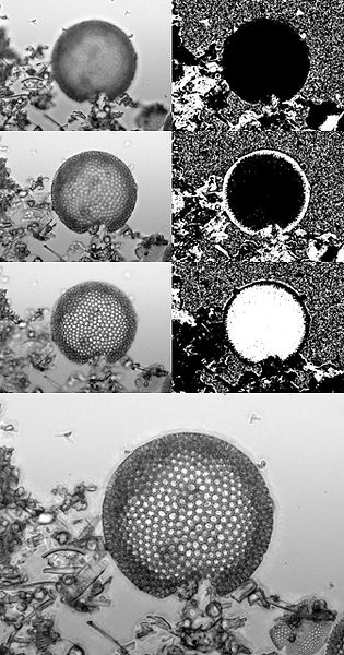
English: Focus stacking (for extended depth of field) in bright field light microscopy. This example is of a diatom microfossil in diatomaceous earth. Top left are the three source image slices at three focal depths. Top right are the contributions (black is no contribution, white is full contribution) of that focal slice to the final "focus stacked" image. Bottom is the resulting focus stacked image with an extended depth of field. Extended depth of field by focus stacking is a powerful tool for light microscopy as at high magnification the depth of field can be extremely shallow, down to around 1 μm. |
| Date | |
| Source | Own work |
| Author | Zephyris |
Licensing
edit
I, the copyright holder of this work, hereby publish it under the following licenses:
This file is licensed under the Creative Commons Attribution-Share Alike 3.0 Unported license.
- You are free:
- to share – to copy, distribute and transmit the work
- to remix – to adapt the work
- Under the following conditions:
- attribution – You must give appropriate credit, provide a link to the license, and indicate if changes were made. You may do so in any reasonable manner, but not in any way that suggests the licensor endorses you or your use.
- share alike – If you remix, transform, or build upon the material, you must distribute your contributions under the same or compatible license as the original.

|
Permission is granted to copy, distribute and/or modify this document under the terms of the GNU Free Documentation License, Version 1.2 or any later version published by the Free Software Foundation; with no Invariant Sections, no Front-Cover Texts, and no Back-Cover Texts. A copy of the license is included in the section entitled GNU Free Documentation License. |
You may select the license of your choice.
File history
Click on a date/time to view the file as it appeared at that time.
| Date/Time | Thumbnail | Dimensions | User | Comment | |
|---|---|---|---|---|---|
| current | 22:15, 13 July 2010 | | 1,344 × 2,560 (986 KB) | Zephyris (talk | contribs) | {{Information |Description={{en|1=Focus stacking (for extended depth of field) in bright field light microscopy. This example is of a diatom microfossil in diatomaceous earth. Top left are the three source image slices at three focal depths. Top right are |
You cannot overwrite this file.
File usage on Commons
There are no pages that use this file.
File usage on other wikis
The following other wikis use this file:
- Usage on en.wikipedia.org
- User talk:Aaadddaaammm
- User talk:Zephyris
- User:Zephyris/Gallery
- Focus stacking
- Talk:Focus stacking
- Wikipedia:Featured pictures/Photographic techniques, terms, and equipment
- Wikipedia:Featured pictures thumbs/26
- Wikipedia:Featured picture candidates/May-2011
- Wikipedia:Featured picture candidates/Focus stacking microscopy
- Wikipedia:Wikipedia Signpost/2011-05-23/Featured content
- Wikipedia:Picture of the day/December 2012
- Template:POTD/2012-12-06
- Wikipedia:Main Page history/2012 December 6
- Wikipedia:Wikipedia Signpost/Single/2011-05-23
- Portal:The arts/Recognized content
- Usage on es.wikipedia.org