File:Habelia fossils details.jpg

Size of this preview: 800 × 534 pixels. Other resolutions: 320 × 214 pixels | 640 × 427 pixels | 1,024 × 684 pixels | 1,280 × 854 pixels | 1,949 × 1,301 pixels.
Original file (1,949 × 1,301 pixels, file size: 840 KB, MIME type: image/jpeg)
Captions
Captions
Add a one-line explanation of what this file represents
Summary edit
| Description |
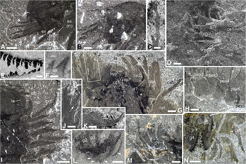
English: Anatomical and morphological details of Habelia optata, morphs A (b, d, f, n) and B (a, e, g, i, l, m). a USNM 139209, close-up of anterior cephalic area, showing intermediary appendage. b USNM 268931, cephalon, showing superimposed insertion of endopods on gnathobases; star points to insertion of anterior endopods. c ROMIP 64357, close-up of fourth cephalic exopodial branch, distal portion showing slender podomeres; arrow points to trident of setae at podomere junction. d ROMIP 64358, close-up of anteriormost region, showing mouth opening and first anterior pairs of gnathobases. e ROMIP 64360, close-up of teeth on masticatory margin of gnathobase; note heavy concentration of carbon in teeth. f Close-up of teeth on masticatory margin of posterior gnathobase on same specimen as in D, showing stronger carbon content in dental edge. g ROMIP 64364, specimen preserved in ventral aspect, close-up of anterior region showing labrum, eyes and appendages; star marks attachment of fifth spinose endopod; arrow points at ornamental spine of cephalic pleura; insets as indicated. h ROMIP 64362, close-up of posterior trunk exopods. i ROMIP 64363, close-up of anterior right cephalic region, dorsal view showing labrum and appendages; arrows point to overprint of gnathobases underneath cephalon. j, k ROMIP 64364. j Close-up of distal portion of cephalic endopod, showing “platform” with setal brushes. k Close-up of terminal claw; arrows point to teeth on inner margin of claw. l USNM 144907, close-up of cephalic gnathobases; arrows point to dentate margins of opposing gnathobases. m ROMIP 64357, close-up on anterior left cephalic region, showing appendages; arrow points to anterior insertion of fourth cephalic endopod. n ROMIP 64359, close-up of cephalic appendages showing insertion of endopods on gnathobases; star marks attachment of fourth cephalic endopod on its gnathobase. c-f, j and k are SEM images; all other are stereomicroscope images of dry specimens under cross-polarized lighting. For abbreviations, see Methods. Scale bars: (a, g, h, i, l, n), 1 mm; (b, m), 0.5 mm; (c, d, k), 200 μm; (e), 100 μm; (f), 50 μm; (j), 500 μm |
| Date | |
| Source | Aria, C., Caron, JB. Mandibulate convergence in an armoured Cambrian stem chelicerate. BMC Evol Biol 17, 261 (2017). https://doi.org/10.1186/s12862-017-1088-7 |
| Author | Cédric Aria & Jean-Bernard Caron |
Licensing edit
This file is licensed under the Creative Commons Attribution 4.0 International license.
- You are free:
- to share – to copy, distribute and transmit the work
- to remix – to adapt the work
- Under the following conditions:
- attribution – You must give appropriate credit, provide a link to the license, and indicate if changes were made. You may do so in any reasonable manner, but not in any way that suggests the licensor endorses you or your use.
File history
Click on a date/time to view the file as it appeared at that time.
| Date/Time | Thumbnail | Dimensions | User | Comment | |
|---|---|---|---|---|---|
| current | 15:36, 10 April 2023 | | 1,949 × 1,301 (840 KB) | Iezer (talk | contribs) | Uploaded a work by Cédric Aria & Jean-Bernard Caron from Aria, C., Caron, JB. Mandibulate convergence in an armoured Cambrian stem chelicerate. BMC Evol Biol 17, 261 (2017). https://doi.org/10.1186/s12862-017-1088-7 with UploadWizard |
You cannot overwrite this file.
File usage on Commons
There are no pages that use this file.