File:Illustration of sacroiliac joint.webp

Size of this PNG preview of this WEBP file: 472 × 599 pixels. Other resolutions: 189 × 240 pixels | 378 × 480 pixels | 896 × 1,137 pixels.
Original file (896 × 1,137 pixels, file size: 66 KB, MIME type: image/webp)
Captions
Captions
Illustration of sacroiliac joint
Summary
edit
| Description |
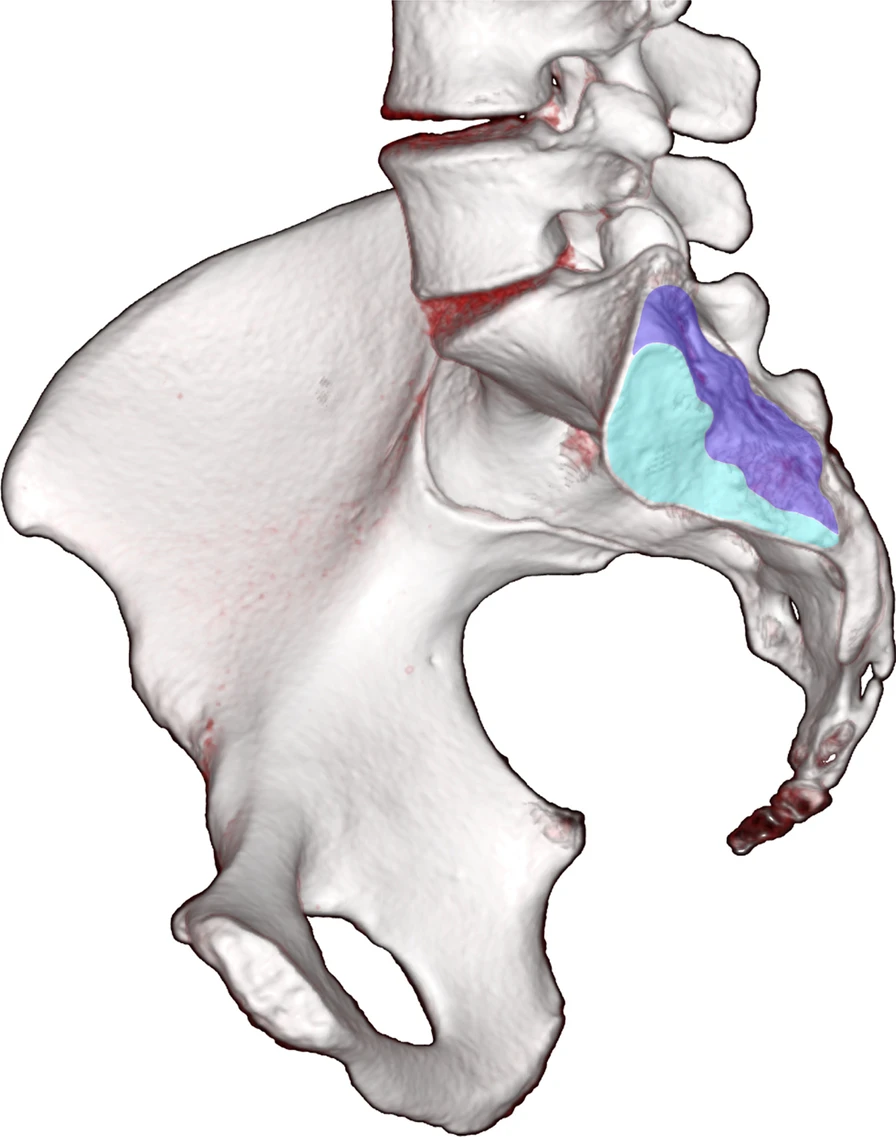
English: Illustration of sacroiliac joint. In this illustration, the sacroiliac joint is divided into an anterior cartilaginous part (light blue) and a more posteriorly located ligamentous part (dark blue) |
| Date | |
| Source | Vereecke, E., Morbée, L., Laloo, F. et al. Anatomical variation of the sacroiliac joints: an MRI study with synthetic CT images. Insights Imaging 14, 30 (2023). https://doi.org/10.1186/s13244-023-01373-1 |
| Author | Vereecke, E., Morbée, L., Laloo, F. et al. |
Licensing
edit
This file is licensed under the Creative Commons Attribution 4.0 International license.
- You are free:
- to share – to copy, distribute and transmit the work
- to remix – to adapt the work
- Under the following conditions:
- attribution – You must give appropriate credit, provide a link to the license, and indicate if changes were made. You may do so in any reasonable manner, but not in any way that suggests the licensor endorses you or your use.
File history
Click on a date/time to view the file as it appeared at that time.
| Date/Time | Thumbnail | Dimensions | User | Comment | |
|---|---|---|---|---|---|
| current | 17:42, 8 March 2024 | | 896 × 1,137 (66 KB) | Balkanique (talk | contribs) | Uploaded a work by Vereecke, E., Morbée, L., Laloo, F. et al. from Vereecke, E., Morbée, L., Laloo, F. et al. Anatomical variation of the sacroiliac joints: an MRI study with synthetic CT images. Insights Imaging 14, 30 (2023). https://doi.org/10.1186/s13244-023-01373-1 with UploadWizard |
You cannot overwrite this file.
File usage on Commons
There are no pages that use this file.