File:Interleukin-1-receptor+IL-1beta 1ITB.png

Size of this preview: 800 × 476 pixels. Other resolutions: 320 × 190 pixels | 640 × 381 pixels | 1,024 × 609 pixels | 1,280 × 762 pixels | 2,027 × 1,206 pixels.
Original file (2,027 × 1,206 pixels, file size: 998 KB, MIME type: image/png)
Captions
Captions
Add a one-line explanation of what this file represents
| Description |
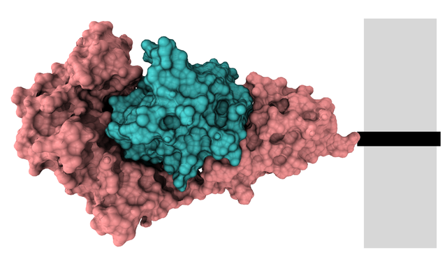
English: Surface model of extracellular interleukin-1 receptor+interleukin-1-beta complex (IL1R pink + IL1B cyan) after PDB 1ITB. The receptor protein continues at the black rectangle into the membrane (grey). Ref.: Vigers GP, Anderson LJ, Caffes P, Brandhuber BJ (March 1997). "Crystal structure of the type-I interleukin-1 receptor complexed with interleukin-1beta". Nature 386 (6621): 190–4. DOI:10.1038/386190a0. PMID 9062193.
This image was created with VMD. |
||
| Date | |||
| Source | Own work | ||
| Author | Ayacop | ||
| Permission (Reusing this file) |
|
| Annotations | This image is annotated: View the annotations at Commons |
File history
Click on a date/time to view the file as it appeared at that time.
| Date/Time | Thumbnail | Dimensions | User | Comment | |
|---|---|---|---|---|---|
| current | 11:22, 1 December 2009 | | 2,027 × 1,206 (998 KB) | Ayacop (talk | contribs) | Different transmembrane position, schematic membrane |
| 10:27, 1 December 2009 |  | 1,878 × 1,329 (1.2 MB) | Ayacop (talk | contribs) | {{Information |Description={{en|1=Surface model of extracellular interleukin-1 receptor+interleukin-1-beta complex (IL1R pink + IL1B cyan) after PDB 1ITB. The receptor continues at the yellow residue into the membrane. Ref.: {{cite journal |author=Vigers |
You cannot overwrite this file.
File usage on Commons
There are no pages that use this file.
File usage on other wikis
The following other wikis use this file:
- Usage on cs.wikipedia.org
- Usage on de.wikipedia.org
- Usage on es.wikipedia.org