File:Isolation and characterization of candidate human ovarian tumor cancer initiating cells form self-renewing, anchorage-independent spheroids under stem cell–selective conditions..jpg
Size of this preview: 306 × 600 pixels. Other resolution: 319 × 625 pixels.
Original file (319 × 625 pixels, file size: 87 KB, MIME type: image/jpeg)
Captions
Captions
Add a one-line explanation of what this file represents
| Description |
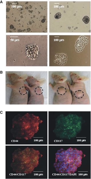
English: (A) Cell suspensions form small, non-adherent clusters 1 wk after plating (top left). After ~10 passages, 1% of spheres persist as larger, symmetric, prototypical spheroids (top right). Typical spheroids contained ~100 cells and could be serially passaged for >6 mo (bottom left). Under differentiating conditions, sphere-forming cells adhere to plates and form symmetric holoclones (bottom right). (B) Injection of ~100 OCICs per mouse from patient tumor 1 (left) or patient tumor 2 (right) dissociated spheroids generated xenograft tumors with 2/2 efficiency. (C) Staining of anti-CD44 monoclonal antibodies (red) in ovarian tumor spheroid (top left); immunofluorescence staining of anti-CD117 monoclonal antibodies (green) in ovarian tumor sphere cells (top right); CD44+ sphere cells colocalize with CD117+ cells (orange overlay, bottom left) and additionally stained with DAPI (blue; bottom right). Magnification, 200x. (Adapted with permission from Zhang, S., Balch, C., Chan, M.W., Lai, H.C., Matei, D., Schilder, J.M., Yan, P.S., Huang, T.H., Nephew, K.P. (2008). Identification and characterization of ovarian cancer-initiating cells from primary human tumors. Cancer Res 68, 4311–4320. Figures 1A, 3A and C.). |
| Date | Published April 30, 2009. |
| Source |
[1] Direct
|
| Author | Chang, H.L., MacLaughlin, D.T., and Donahoe, P.K., Somatic stem cells of the ovary and their relationship to human ovarian cancers (April 30, 2009), StemBook, ed. The Stem Cell Research Community, StemBook, doi/10.3824/stembook.1.43.1, http://www.stembook.org. |
| Permission (Reusing this file) |
This file is licensed under the Creative Commons Attribution 3.0 Unported license.
|
File history
Click on a date/time to view the file as it appeared at that time.
| Date/Time | Thumbnail | Dimensions | User | Comment | |
|---|---|---|---|---|---|
| current | 18:45, 5 April 2013 | | 319 × 625 (87 KB) | Smallbot (talk | contribs) | Uploading CC-BY images from the the StemBook http://www.stembook.org/ 42/173 |
You cannot overwrite this file.
File usage on Commons
There are no pages that use this file.