File:JCS045203F1.jpg
No higher resolution available.
JCS045203F1.jpg (439 × 435 pixels, file size: 122 KB, MIME type: image/jpeg)
Captions
Captions
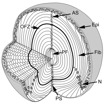
Mouse eye lens drawing showing main features of ultrastucture through cutout 3D.
Summary
edit
| Description |
English: Cellular organization of the mouse lens. The lens consists of two cell types: epithelial cells (Epi) located at the anterior surface, and fiber cells (Fib), which comprise the remainder and majority of the tissue. At the lens equator, epithelial cells differentiate into fiber cells. As they differentiate, fibers become highly elongated. The tips of the elongating fibers converge at the anterior and posterior lens sutures (AS and PS, respectively). In cross section, the fiber cells have a flattened hexagonal profile with two broad faces (oriented parallel to the lens surface) and four narrow faces. Initially, all fiber cells are nucleated but, during differentiation, nuclei (N) and other organelles are degraded. As a result, the central region of the lens constitutes an organelle-free zone (OFZ). The innermost cells, termed primary fiber cells (PF), are formed during embryonic development and are less regular in shape and arrangement than the other fiber cells. |
| Date | |
| Source |
J Cell Sci. 2009 May 15;122(Pt 10):1607-15. doi: 10.1242/jcs.045203. Epub 2009 Apr 28. The stratified syncytium of the vertebrate lens by Yanrong Shi 1 , Kelly Barton, Alicia De Maria, J Mark Petrash, Alan Shiels, Steven Bassnett PMID: 19401333 PMCID: PMC2680101 DOI: 10.1242/jcs.045203 |
| Author | Yanrong Shi 1 , Kelly Barton, Alicia De Maria, J Mark Petrash, Alan Shiels, Steven Bassnett |
Licensing
edit
Pubmed central open access article already reused and adapted in other open access articles
This file is licensed under the Creative Commons Attribution 4.0 International license.
- You are free:
- to share – to copy, distribute and transmit the work
- to remix – to adapt the work
- Under the following conditions:
- attribution – You must give appropriate credit, provide a link to the license, and indicate if changes were made. You may do so in any reasonable manner, but not in any way that suggests the licensor endorses you or your use.
. Right to use confirmed by email from artist Steven Bassnett 12:06pm Jan 26th 2023.
File history
Click on a date/time to view the file as it appeared at that time.
| Date/Time | Thumbnail | Dimensions | User | Comment | |
|---|---|---|---|---|---|
| current | 08:54, 26 January 2023 | | 439 × 435 (122 KB) | Tgru001 (talk | contribs) | Uploaded a work by Yanrong Shi 1 , Kelly Barton, Alicia De Maria, J Mark Petrash, Alan Shiels, Steven Bassnett from J Cell Sci. 2009 May 15;122(Pt 10):1607-15. doi: 10.1242/jcs.045203. Epub 2009 Apr 28. The stratified syncytium of the vertebrate lens by Yanrong Shi 1 , Kelly Barton, Alicia De Maria, J Mark Petrash, Alan Shiels, Steven Bassnett PMID: 19401333 PMCID: PMC2680101 DOI: 10.1242/jcs.045203 with UploadWizard |
You cannot overwrite this file.
File usage on Commons
The following page uses this file: