File:Journal.ppat.1005203.g001.png
Size of this preview: 661 × 600 pixels. Other resolutions: 265 × 240 pixels | 529 × 480 pixels | 847 × 768 pixels | 1,129 × 1,024 pixels | 2,221 × 2,015 pixels.
Original file (2,221 × 2,015 pixels, file size: 3.61 MB, MIME type: image/png)
Captions
Captions
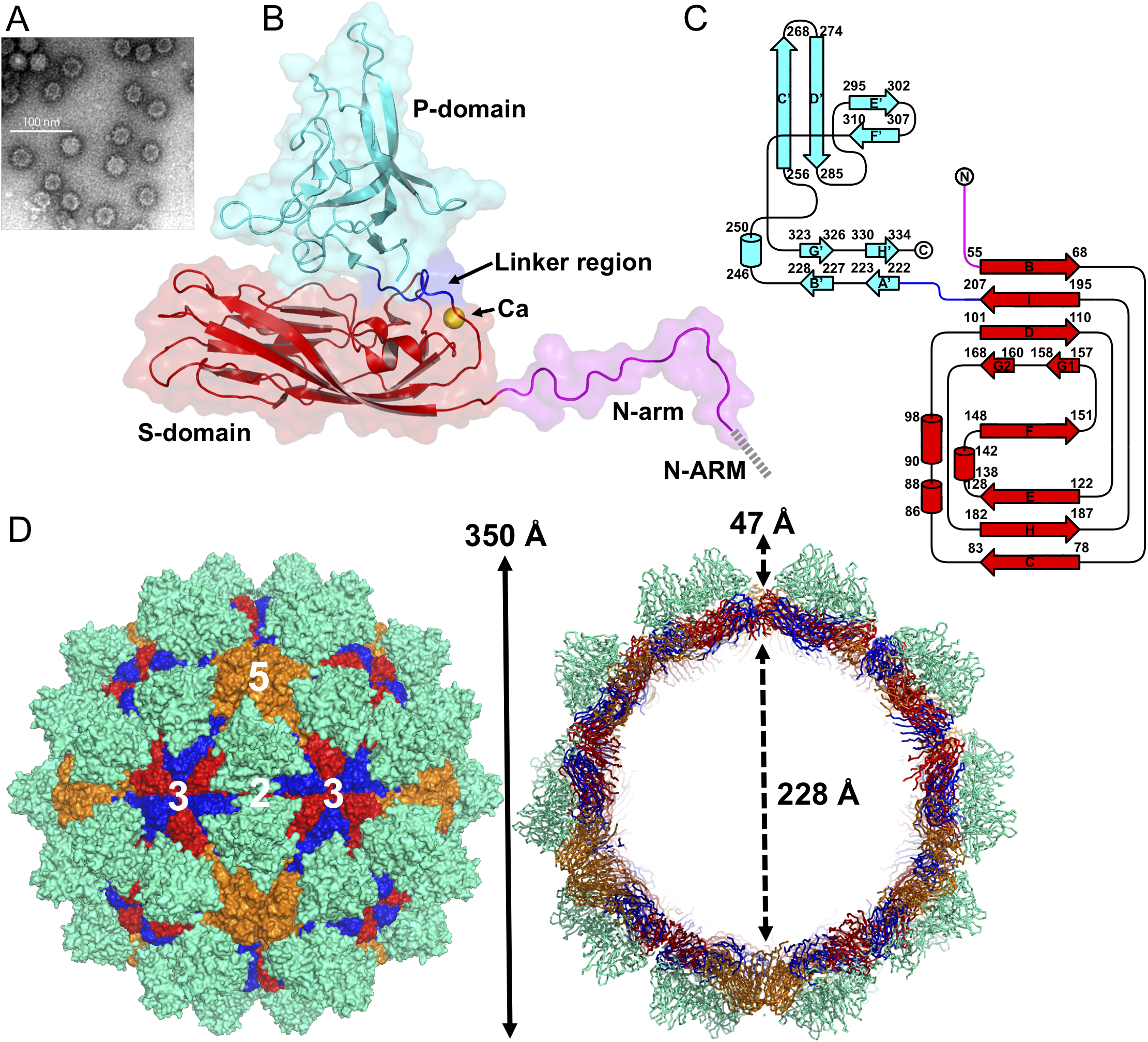
EM image and the overall structure of grouper nervous necrosis virus (GNNV).
Summary
edit
| Description |
English: EM image and the overall structure of grouper nervous necrosis virus (GNNV). (A) A representative negative-staining EM image of the purified GNNV-LPs after self-assembly. (B) A ribbon presentation of the subunit C of GNNV-LP. The disordered N-ARM (residue 1−33, gray), N-arm (residues 34−51, magenta), the S-domain (residues 52−213, red), the linker region (residues 214−220, blue), the P-domain (residues 221−338, cyan) and Ca2+ ion (yellow sphere) are shown. (C) A topology diagram of GNNV CP with the helices and strands in cylinders and arrows, respectively. The 1D topology of the subunit C is color-coded as in B. (D) Surface domain-colored diagram (left) and central cavity (right) representations of the T = 3 GNNV-LP. The tip-to-tip distance is ~350 Å, the diameter of the central cavity is ~228 Å, and the spike protrusion on the capsid surface is ~47 Å. The S-domains of the subunits A, B and C are shown in orange, blue and red, respectively, and the P-domains are shown in cyan. The structure of the GNNV-LP is viewed along the I2, I3 and I5 axes. |
| Date | |
| Source | Chen N-C, Yoshimura M, Guan H-H, Wang T-Y, Misumi Y, Lin C-C, et al. (2015) Crystal Structures of a Piscine Betanodavirus: Mechanisms of Capsid Assembly and Viral Infection. PLoS Pathog 11(10): e1005203. https://doi.org/10.1371/journal.ppat.1005203 (Fig. 1) |
| Author | Nai-Chi Chen, Masato Yoshimura, Hong-Hsiang Guan, Ting-Yu Wang, Yuko Misumi, Chien-Chih Lin, Phimonphan Chuankhayan, Atsushi Nakagawa, Sunney I. Chan, Tomitake Tsukihara, Tzong-Yueh Chen, Chun-Jung Chen |
| Other versions |
  |
Licensing
edit
This file is licensed under the Creative Commons Attribution 4.0 International license.
- You are free:
- to share – to copy, distribute and transmit the work
- to remix – to adapt the work
- Under the following conditions:
- attribution – You must give appropriate credit, provide a link to the license, and indicate if changes were made. You may do so in any reasonable manner, but not in any way that suggests the licensor endorses you or your use.
File history
Click on a date/time to view the file as it appeared at that time.
| Date/Time | Thumbnail | Dimensions | User | Comment | |
|---|---|---|---|---|---|
| current | 11:58, 6 December 2020 | | 2,221 × 2,015 (3.61 MB) | Guest2625 (talk | contribs) | Uploaded a work by Nai-Chi Chen, Masato Yoshimura, Hong-Hsiang Guan, Ting-Yu Wang, Yuko Misumi, Chien-Chih Lin, Phimonphan Chuankhayan, Atsushi Nakagawa, Sunney I. Chan, Tomitake Tsukihara, Tzong-Yueh Chen, Chun-Jung Chen from Chen N-C, Yoshimura M, Guan H-H, Wang T-Y, Misumi Y, Lin C-C, et al. (2015) Crystal Structures of a Piscine Betanodavirus: Mechanisms of Capsid Assembly and Viral Infection. PLoS Pathog 11(10): e1005203. https://doi.org/10.1371/journal.ppat.1005203 with UploadWizard |
You cannot overwrite this file.
File usage on Commons
The following 2 pages use this file:
File usage on other wikis
The following other wikis use this file:
- Usage on en.wikipedia.org