File:Micrograph of atypical ductal hyperplasia.jpg
Size of this preview: 456 × 600 pixels. Other resolutions: 182 × 240 pixels | 593 × 780 pixels.
Original file (593 × 780 pixels, file size: 219 KB, MIME type: image/jpeg)
Captions
Captions
Micrograph of atypical ductal hyperplasia
Summary
edit
| Description |
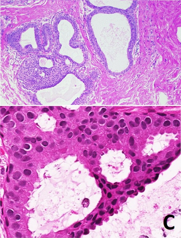
English: Histological appearance of atypical ductal hyperplasia (ADH) and immunohistochemical phenotype. - Top image - One focus (< 2 mm) of two architecturally disarranged cross sections of tubuli showing a monotonous intraductal proliferation with secondary intraluminal architecture. Hematoxylin and Eosin stain. - Bottom image - Higher magnification of ADH shows low-grade nuclear atypia and monotonous cell proliferation along with secondary intraluminal architecture. Hematoxylin and Eosin stain. |
| Date | |
| Source |
(2018). "Atypical ductal hyperplasia and the risk of underestimation: tissue sampling method, multifocality, and associated calcification significantly influence the diagnostic upgrade rate based on subsequent surgical specimens". Breast Cancer 26 (4): 452–458. DOI:10.1007/s12282-018-00943-2. ISSN 1340-6868.
|
| Author | Christoph J. Rageth, Ravit Rubenov, Cristian Bronz, Daniel Dietrich, Christoph Tausch, Ann-Katrin Rodewald, Zsuzsanna Varga |
| Other versions |
 |
Licensing
edit
This file is licensed under the Creative Commons Attribution 4.0 International license.
- You are free:
- to share – to copy, distribute and transmit the work
- to remix – to adapt the work
- Under the following conditions:
- attribution – You must give appropriate credit, provide a link to the license, and indicate if changes were made. You may do so in any reasonable manner, but not in any way that suggests the licensor endorses you or your use.
File history
Click on a date/time to view the file as it appeared at that time.
| Date/Time | Thumbnail | Dimensions | User | Comment | |
|---|---|---|---|---|---|
| current | 11:24, 2 October 2019 | | 593 × 780 (219 KB) | Mikael Häggström (talk | contribs) | User created page with UploadWizard |
You cannot overwrite this file.
File usage on Commons
The following 2 pages use this file: