File:Nicotiana tabacum fpls-12-642879-g001.jpg

Size of this preview: 451 × 599 pixels. Other resolutions: 180 × 240 pixels | 361 × 480 pixels | 578 × 768 pixels | 770 × 1,024 pixels | 2,126 × 2,826 pixels.
Original file (2,126 × 2,826 pixels, file size: 878 KB, MIME type: image/jpeg)
Captions
Captions
Add a one-line explanation of what this file represents
Summary edit
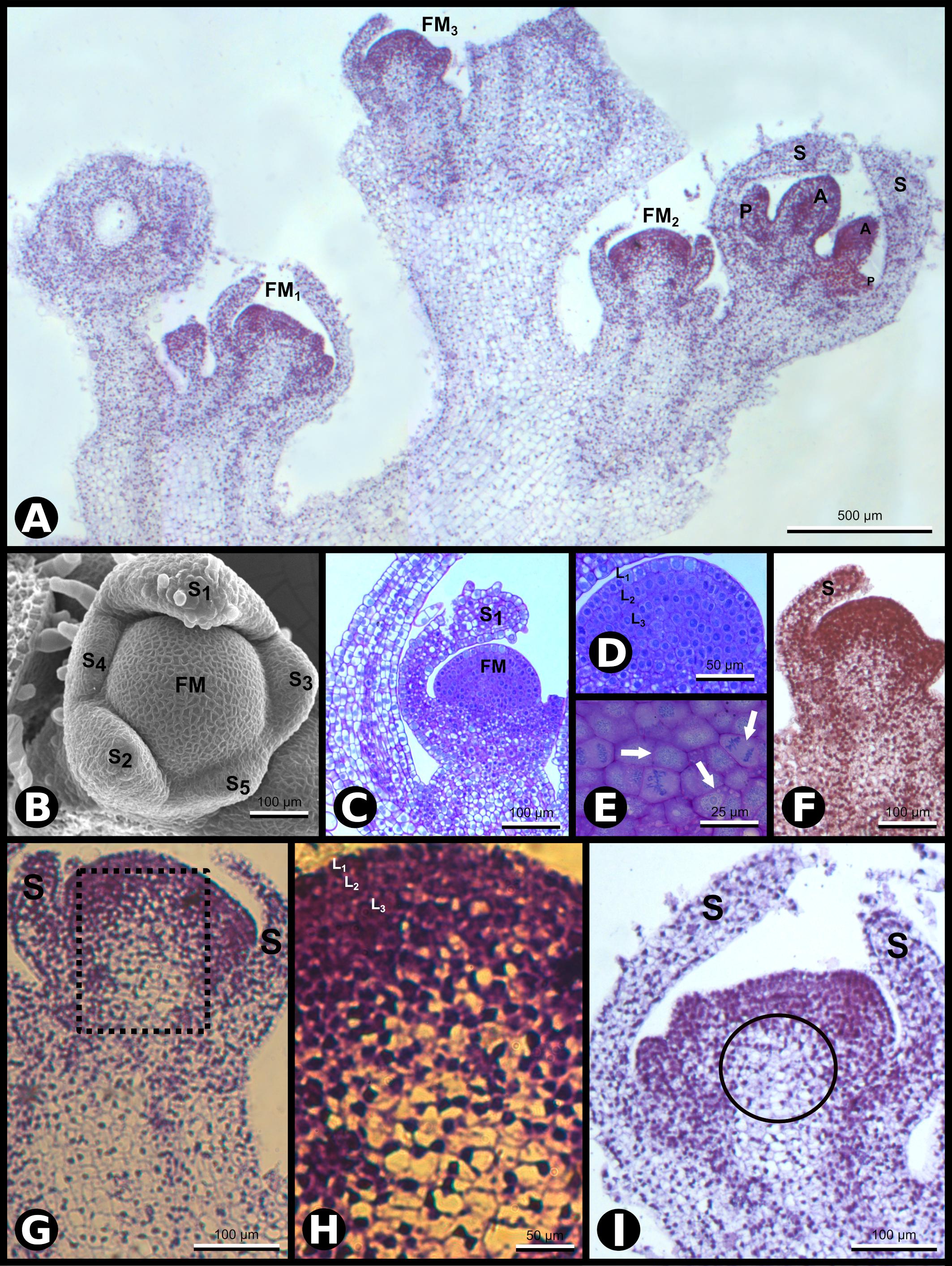
| Description | Figure 1. SCI1 expression during Nicotiana tabacum early floral development. (A) In situ hybridization of inflorescence apex with SCI1 antisense probe. Four floral buds are observed. Scale bar: 500 μm. (B) Scanning electron microscopy (SEM) showing the asynchronous emergence of the sepals (S1–S5) in a flower meristem and sepal S1 with trichomes (stage –9, here defined). Scale bar: 100 μm. (C) Bright-field microscopy showing a longitudinal section of a very young flower bud at stage –9. Scale bar: 100 μm. (D) A higher magnification view of the flower bud in C, in which the three meristematic cell layers (L1, L2, and L3) are seen. Scale bar: 50 μm. (E) A higher magnification view of the flower meristem shown in (D), in which cell divisions are visible (arrows). Scale bar: 25 μm. (F–I) In situ hybridization with SCI1 antisense probe of very young flower buds, even before stage –7, the youngest developmental stage defined by Koltunow et al. (1990). (F) Flower meristem with emerging sepals (stage –10, here defined). This is a higher magnification of FM3 from (A). Scale bar: 100 μm. (G) Flower meristem with sepal primordia (stage –9, here defined). This is a higher magnification of FM2 from (A). Scale bar: 100 μm. (H) A higher magnification view of the marked area in (G). The meristematic cell layers (L1, L2, and L3) are identifiable. Scale bar: 50 μm. (I) Flower meristem with emerging petals and anthers, at stage –8 (here defined). This is a higher magnification of FM1 from (A). Scale bar: 100 μm. Floral meristem (FM), sepals (S). Compare the image shown in (F) with the images (G–I) and observe the reduced SCI1 expression in the OC [encircled in (I)]. |
| Date | 18-03-2021 |
| Source | https://www.frontiersin.org/files/Articles/642879/fpls-12-642879-HTML/image_m/fpls-12-642879-g001.jpg, SCI1 Is a Direct Target of AGAMOUS and WUSCHEL and Is Specifically Expressed in the Floral Meristematic Cells, Front. Plant Sci., 18 March 2021,Sec. Plant Development and EvoDevo, Volume 12 - 2021, https://doi.org/10.3389/fpls.2021.642879 |
| Author | Joelma O. Cruz Juca A. B. San Martin, Greice Lubini, Edward J. Strini1, Rómulo Sobral, Vitor F. Pinoti, Pedro B. Ferreira, Vanessa Thomé, Andréa C. Quiapim, Marcelo C. Dornelas, Maria Cristina, S. Pranchevicius, Francisco Madueño, M. Manuela R. Costa, Maria Helena S. Goldman, |
Open access
Licensing edit
This file is licensed under the Creative Commons Attribution 4.0 International license.
- You are free:
- to share – to copy, distribute and transmit the work
- to remix – to adapt the work
- Under the following conditions:
- attribution – You must give appropriate credit, provide a link to the license, and indicate if changes were made. You may do so in any reasonable manner, but not in any way that suggests the licensor endorses you or your use.
File history
Click on a date/time to view the file as it appeared at that time.
| Date/Time | Thumbnail | Dimensions | User | Comment | |
|---|---|---|---|---|---|
| current | 20:32, 29 December 2023 | | 2,126 × 2,826 (878 KB) | Rasbak (talk | contribs) | {{Information |description=Figure 1. SCI1 expression during Nicotiana tabacum early floral development. (A) In situ hybridization of inflorescence apex with SCI1 antisense probe. Four floral buds are observed. Scale bar: 500 μm. (B) Scanning electron microscopy (SEM) showing the asynchronous emergence of the sepals (S1–S5) in a flower meristem and sepal S1 with trichomes (stage –9, here defined). Scale bar: 100 μm. (C) Bright-field microscopy showing a longitudinal section of a very young flo... |
You cannot overwrite this file.
File usage on Commons
There are no pages that use this file.
File usage on other wikis
The following other wikis use this file:
- Usage on nl.wikipedia.org