File:Nicotiana tabacum fpls-12-642879-g002.jpg

Size of this preview: 497 × 599 pixels. Other resolutions: 199 × 240 pixels | 398 × 480 pixels | 637 × 768 pixels | 849 × 1,024 pixels | 2,126 × 2,564 pixels.
Original file (2,126 × 2,564 pixels, file size: 1.06 MB, MIME type: image/jpeg)
Captions
Captions
Add a one-line explanation of what this file represents
Summary edit
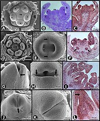
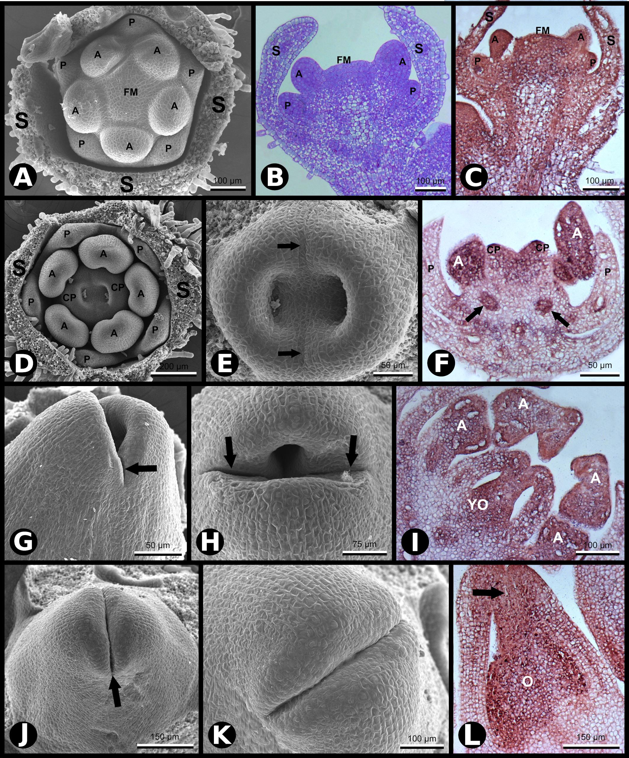
| Description | Figure 2. SCI1 expression during later stages of floral development (continuation of the stages shown in Figure 1). (A) SEM of a flower bud in which petals and anthers are emerging (advanced stage –8). Scale bar: 100 μm. (B) Bright-field microscopy showing a longitudinal section of a flower bud in a developmental stage equivalent to the one shown in (A). Scale bar: 100 μm. (C) In situ hybridization of a flower bud (advanced stage –8) with SCI1 antisense probe. Scale bar: 100 μm. (D) SEM of a flower bud at stage –7 (as defined by Koltunow et al., 1990), in which carpels are emerging. Scale bar: 200 μm. (E) A higher magnification view of the flower bud shown in (D), in which the fusion lines are visible (arrows). Scale bar: 50 μm. (F) In situ hybridization of a flower bud at stage –7/–6, with SCI1 antisense probe. Arrows point to ovary locules. Scale bar: 50 μm. (G,H) SEM of flower buds at stage –6; carpels fused at the base and not yet fused at the top. Scale bars: 50 μm (G) and 75 μm (H). (I) In situ hybridization of a flower bud at late stage –6, with SCI1 antisense probe. Scale bar: 100 μm. (J,K) SEM of flower buds at stage –5; carpels already fused at the top; the fusion region is a site of intense cell proliferation. Scale bars: 150 μm (J) and 100 μm (K). (L) In situ hybridization with SCI1 antisense probe of a flower bud at late stage –5; style beginning to form (arrow). Scale bar: 150 μm. Floral meristem (FM), sepals (S), petals (P), anther (A), carpels (C), carpel primordia (CP), ovary (O), young ovary (YO). |
| Date | |
| Source | https://www.frontiersin.org/files/Articles/642879/fpls-12-642879-HTML/image_m/fpls-12-642879-g002.jpg, SCI1 Is a Direct Target of AGAMOUS and WUSCHEL and Is Specifically Expressed in the Floral Meristematic Cells, Front. Plant Sci., 18 March 2021,Sec. Plant Development and EvoDevo, Volume 12 - 2021, https://doi.org/10.3389/fpls.2021.642879 |
| Author | Joelma O. Cruz Juca A. B. San Martin, Greice Lubini, Edward J. Strini1, Rómulo Sobral, Vitor F. Pinoti, Pedro B. Ferreira, Vanessa Thomé, Andréa C. Quiapim, Marcelo C. Dornelas, Maria Cristina, S. Pranchevicius, Francisco Madueño, M. Manuela R. Costa, Maria Helena S. Goldman, |
Open access
Licensing edit
This file is licensed under the Creative Commons Attribution 4.0 International license.
- You are free:
- to share – to copy, distribute and transmit the work
- to remix – to adapt the work
- Under the following conditions:
- attribution – You must give appropriate credit, provide a link to the license, and indicate if changes were made. You may do so in any reasonable manner, but not in any way that suggests the licensor endorses you or your use.
File history
Click on a date/time to view the file as it appeared at that time.
| Date/Time | Thumbnail | Dimensions | User | Comment | |
|---|---|---|---|---|---|
| current | 22:28, 29 December 2023 | | 2,126 × 2,564 (1.06 MB) | Rasbak (talk | contribs) | {{Information |description=Figure 2. SCI1 expression during later stages of floral development (continuation of the stages shown in Figure 1). (A) SEM of a flower bud in which petals and anthers are emerging (advanced stage –8). Scale bar: 100 μm. (B) Bright-field microscopy showing a longitudinal section of a flower bud in a developmental stage equivalent to the one shown in (A). Scale bar: 100 μm. (C) In situ hybridization of a flower bud (advanced stage –8) with SCI1 antisense probe. Scale... |
You cannot overwrite this file.
File usage on Commons
There are no pages that use this file.
File usage on other wikis
The following other wikis use this file:
- Usage on nl.wikipedia.org