File:PARP1 binding olaparib 5DS3.png

Size of this preview: 715 × 599 pixels. Other resolutions: 286 × 240 pixels | 573 × 480 pixels | 916 × 768 pixels | 1,222 × 1,024 pixels | 1,440 × 1,207 pixels.
Original file (1,440 × 1,207 pixels, file size: 1.12 MB, MIME type: image/png)
Captions
Captions
Add a one-line explanation of what this file represents
Summary edit
| Description |
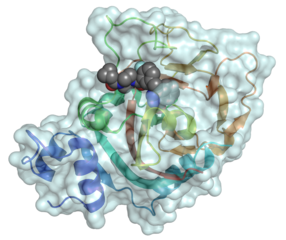
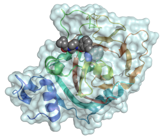
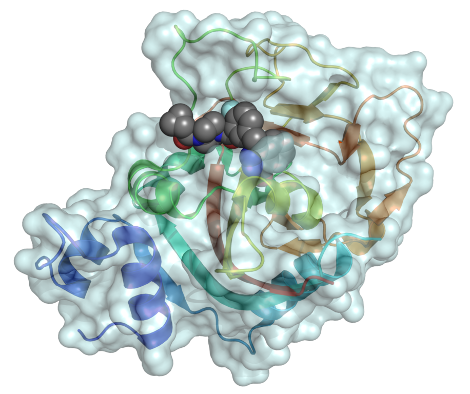
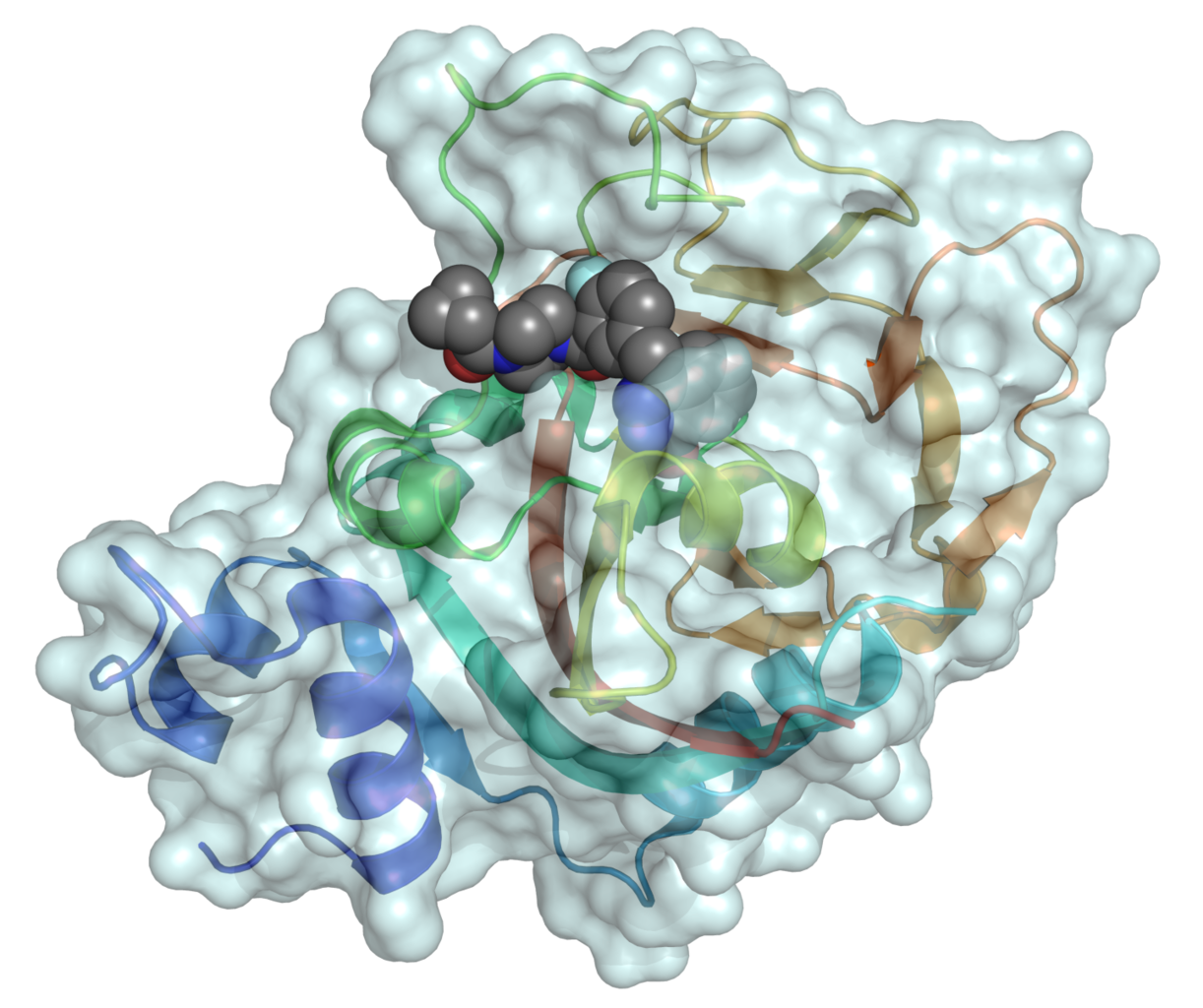
Mixed surface–ribbon representation of the catalytic domain of human poly (ADP-ribose) polymerase 1 (PARP1) binding the small-molecule inhibitor olaparib (shown as a space-filling model). Representation-independent color control was used to color the surface and ribbon diagrams differentially in the interest of clarity. This structure was created with PyMOL. |
||
| Date | |||
| Source |
From PDB entry 5DS3. More information:
|
||
| Author | Fvasconcellos (talk · contribs) | ||
| Permission (Reusing this file) |
|
File history
Click on a date/time to view the file as it appeared at that time.
| Date/Time | Thumbnail | Dimensions | User | Comment | |
|---|---|---|---|---|---|
| current | 04:54, 11 August 2017 | | 1,440 × 1,207 (1.12 MB) | Fvasconcellos (talk | contribs) | =={{int:filedesc}}== {{Information |Description=Mixed surface–ribbon representation of the {{w|catalytic domain}} of human {{w|poly (ADP-ribose) polymerase}} 1 ({{w|PARP1}}) binding the small-molecule inhibitor {{w|olaparib}} (shown as a {{w|space-fi... |
You cannot overwrite this file.
File usage on Commons
There are no pages that use this file.
File usage on other wikis
The following other wikis use this file:
- Usage on en.wikipedia.org
- Usage on pl.wikipedia.org