File:Paracentrotus lividus anatomy of early and late juveniles.jpg

Size of this preview: 533 × 600 pixels. Other resolutions: 213 × 240 pixels | 426 × 480 pixels | 682 × 768 pixels | 1,117 × 1,257 pixels.
Original file (1,117 × 1,257 pixels, file size: 2.01 MB, MIME type: image/jpeg)
Captions
Captions
Add a one-line explanation of what this file represents
Summary
edit
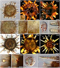
| Description | FIGURE 16. Anatomy of early and late juveniles of Paracentrotus lividus. Developmental stages are as follows: (A–I) early juvenile stage, 1 day post-metamorphosis (EJuv); (J–Q) late juvenile stage, 8 days post-metamorphosis (LJuv). In (A,D–J,M–Q), images were acquired using bright-field light microscopy. In (B,C,K,L), images were obtained using polarized light microscopy. In (A–D,J–L), images correspond to specimens in oral view in (A,C,D,J,L) and in aboral view in (B,K). ((B) inset) Close-up of an opened pedicellaria. ((C) inset) Close-up of the skeletal elements constituting the primordia of the adult masticatory apparatus. (D) Close-up of the ring and radial canals of the water vascular system. (E) Close-up of a pair of growing secondary podia located underneath a primary podium. (F) Close-up of the ampullae formed at the proximal tip of a radial canal. (G) Close-up of the skeletal disk within the papilla of a primary podium. (H) Close-up of a developing definitive spine. (I) Close-up of a developing juvenile spine. (M) Close-up of a sphaeridium. (N) Close-up of a degenerated primary podium. (O) Close-up of the skeletal disk within the papilla of a secondary podium. (P) Close-up of a fully formed definitive spine. (Q) Close-up of a fully formed juvenile spine. In (B), cyan asterisks mark the pedicellariae present on the aboral surface of the juvenile, and white dotted lines delineate the genital plates. In ((C) inset), the white arrowhead indicates a tooth primordium, cyan arrowheads highlight primordia of the hemipyramids, and green arrowheads mark primordia of the epiphyses. In (F), white dotted lines outline two ampullae formed at the proximal tip of a radial canal. In (H,I), white arrowheads indicate the apex of a growing definitive spine in (H) and of a growing juvenile spine in (I). Scale bar: (A–C,J–L) 100 μm; ((B) inset, (C) inset, (D–I,M–Q)) 30 µm. Amp: ampulla; AmbP: ambulacral plate; BucP: buccal plate; DS: definitive spine; gAB: genital plate AB; gBC: genital plate BC; gCD: genital plate CD; gDE: genital plate DE; gEA: genital plate EA; JS: juvenile spine; Peri: peristome; PP: primary podium; RaC: radial canal; RiC: ring canal; SP: secondary podium; Sph: sphaeridium; SR: skeletal rod; Tub: tubercule. |
| Date | |
| Source |
https://www.frontiersin.org/articles/10.3389/fcell.2022.966408/full Developmental atlas of the indirect-developing sea urchin Paracentrotus lividus: From fertilization to juvenile stages, Front. Cell Dev. Biol., 31 October 2022 Sec. Morphogenesis and Patterning Volume 10 - 2022, https://doi.org/10.3389/fcell.2022.966408 |
| Author | Laurent Formery, Axel Wakefield, Maeva Gesson, Ludovic Toisoul, Guy Lhomond, Laurent Gilletta, Régis Lasbleiz, Michael Schubert, Jenifer C. Croce |
Licensing
edit
This file is licensed under the Creative Commons Attribution 4.0 International license.
- You are free:
- to share – to copy, distribute and transmit the work
- to remix – to adapt the work
- Under the following conditions:
- attribution – You must give appropriate credit, provide a link to the license, and indicate if changes were made. You may do so in any reasonable manner, but not in any way that suggests the licensor endorses you or your use.

|
This file, which was originally posted to an external website, has not yet been reviewed by an administrator or reviewer to confirm that the above license is valid. See Category:License review needed for further instructions.
|
File history
Click on a date/time to view the file as it appeared at that time.
| Date/Time | Thumbnail | Dimensions | User | Comment | |
|---|---|---|---|---|---|
| current | 10:13, 7 March 2024 | | 1,117 × 1,257 (2.01 MB) | Rasbak (talk | contribs) | {{Information |description=FIGURE 16. Anatomy of early and late juveniles of Paracentrotus lividus. Developmental stages are as follows: (A–I) early juvenile stage, 1 day post-metamorphosis (EJuv); (J–Q) late juvenile stage, 8 days post-metamorphosis (LJuv). In (A,D–J,M–Q), images were acquired using bright-field light microscopy. In (B,C,K,L), images were obtained using polarized light microscopy. In (A–D,J–L), images correspond to specimens in oral view in (A,C,D,J,L) and in aboral view in... |
You cannot overwrite this file.
File usage on Commons
The following 2 pages use this file:
File usage on other wikis
The following other wikis use this file:
- Usage on nl.wikipedia.org