File:Paracentrotus lividus development of the pedicellariae and the genital plates.jpg

Size of this preview: 800 × 525 pixels. Other resolutions: 320 × 210 pixels | 640 × 420 pixels | 1,024 × 672 pixels | 1,423 × 934 pixels.
Original file (1,423 × 934 pixels, file size: 1.42 MB, MIME type: image/jpeg)
Captions
Captions
Add a one-line explanation of what this file represents
Summary
edit
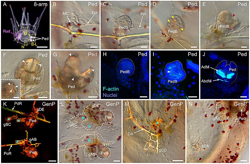
| Description | FIGURE 14. Development of the pedicellariae and the genital plates in Paracentrotus lividus larvae. In (A–N), images are from larvae at the 8-arm pluteus stage (8-arm). In (A), the image was acquired using dark-field light microscopy. In (B–G,L–N), images were obtained using bright-field light microscopy. In (H–J), images are maximum intensity projections of confocal z-stacks of larvae co-labeled for F-actin (muscles; cyan) and DNA (nuclei; blue), and they correspond to projections of the entire pedicellaria. In (K), the image was taken using polarized light to highlight the skeletal elements. In (A), the larva is in anterior view, with the ventral side up and the left side to the left. (B–N) Close-ups of the regions outlined by yellow boxes in (A). (B–J) Close-ups of pedicellariae (Ped). (K–N) Close-ups of genital plates (GenP). In (A), the purple dotted line delineates the adult rudiment on the left side of the larva. In (D), yellow arrowheads indicate the skeletal elements developing inside the pedicellaria bud. In (E), the red arrowhead shows the individualization of the three lobes within the bud. In ((F), (F) inset, (G)), white arrowheads mark the three jaws of the pedicellaria, which can be either closed like in (F) and ((F) inset) or open like in (G). In (H–J), white dotted lines outline the pedicellaria bud in (H–I) or the pedicellaria jaws in (J). In (I), the white arrowhead points to the first muscle fibers appearing within a pedicellaria bud. In (K–M), cyan asterisks indicate the position of juvenile spines associated with the genital plates. In (L), white dotted lines delineate the genital plates and their associated juvenile spines developing in the vicinity of pedicellariae. Scale bar: (A) 200 μm; ((B–G), (F) inset) 15 μm; (H–J,K–M) 30 μm; (N) 50 µm. AdM: adductor muscle; AbdM: abductor muscle; DA: dorsal arch; Epi: larval epidermis; gAB: genital plate AB; gBC: genital plate BC; gCD: genital plate CD; gDE: genital plate DE; gEA: genital plate EA; GenP: genital plate; MC: mesenchyme cell; PdR: posterodorsal rod; Ped: pedicellaria; PedB: pedicellaria bud; PoR: postoral rod; Rud: adult rudiment. |
| Date | |
| Source |
https://www.frontiersin.org/articles/10.3389/fcell.2022.966408/full Developmental atlas of the indirect-developing sea urchin Paracentrotus lividus: From fertilization to juvenile stages, Front. Cell Dev. Biol., 31 October 2022 Sec. Morphogenesis and Patterning Volume 10 - 2022, https://doi.org/10.3389/fcell.2022.966408 |
| Author | Laurent Formery, Axel Wakefield, Maeva Gesson, Ludovic Toisoul, Guy Lhomond, Laurent Gilletta, Régis Lasbleiz, Michael Schubert, Jenifer C. Croce |
Licensing
edit
This file is licensed under the Creative Commons Attribution 4.0 International license.
- You are free:
- to share – to copy, distribute and transmit the work
- to remix – to adapt the work
- Under the following conditions:
- attribution – You must give appropriate credit, provide a link to the license, and indicate if changes were made. You may do so in any reasonable manner, but not in any way that suggests the licensor endorses you or your use.

|
This file, which was originally posted to an external website, has not yet been reviewed by an administrator or reviewer to confirm that the above license is valid. See Category:License review needed for further instructions.
|
File history
Click on a date/time to view the file as it appeared at that time.
| Date/Time | Thumbnail | Dimensions | User | Comment | |
|---|---|---|---|---|---|
| current | 22:11, 6 March 2024 | | 1,423 × 934 (1.42 MB) | Rasbak (talk | contribs) | {{Information |description=FIGURE 14. Development of the pedicellariae and the genital plates in Paracentrotus lividus larvae. In (A–N), images are from larvae at the 8-arm pluteus stage (8-arm). In (A), the image was acquired using dark-field light microscopy. In (B–G,L–N), images were obtained using bright-field light microscopy. In (H–J), images are maximum intensity projections of confocal z-stacks of larvae co-labeled for F-actin (muscles; cyan) and DNA (nuclei; blue), and they correspon... |
You cannot overwrite this file.
File usage on Commons
The following 2 pages use this file:
File usage on other wikis
The following other wikis use this file:
- Usage on nl.wikipedia.org