File:Pedicellina echinata Early stages in the development of the egg.jpg

Size of this preview: 800 × 528 pixels. Other resolutions: 320 × 211 pixels | 640 × 423 pixels | 1,024 × 676 pixels | 1,278 × 844 pixels.
Original file (1,278 × 844 pixels, file size: 239 KB, MIME type: image/jpeg)
Captions
Captions
Add a one-line explanation of what this file represents
Summary edit
| Description |
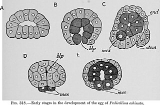
Fig. 318.—Early stages in the development of the egg of Pedicellina echinata. (After Hatschek.) A, flattened blastula. B, gastrula. C, optical section of later stage nearly sagittal Showing the mother cell of the mesoderm of the right side, the endodermic sac, and the stomodaeal invagination. D, view of stage in which the blastopore is closing seen from below. E, frontal section of the same stage; blp, blastopore ; end, endodermic sac ; mes, mother cells of the mesoderm |
| Date | |
| Source | https://www.flickr.com/photos/internetarchivebookimages/20781139341/in/photostream/ Text-book of embryology. London : Macmillan |
| Author | MacBride, E. W.; Kerr, John Graham ; Heape, Walter |
Licensing edit
| This file is made available under the Creative Commons CC0 1.0 Universal Public Domain Dedication. | |
| The person who associated a work with this deed has dedicated the work to the public domain by waiving all of their rights to the work worldwide under copyright law, including all related and neighboring rights, to the extent allowed by law. You can copy, modify, distribute and perform the work, even for commercial purposes, all without asking permission.
|
| This image was originally posted to Flickr by Internet Archive Book Images at https://flickr.com/photos/126377022@N07/20781139341. It was reviewed on 23 March 2024 by FlickreviewR 2 and was confirmed to be licensed under the terms of the cc-zero. |
File history
Click on a date/time to view the file as it appeared at that time.
| Date/Time | Thumbnail | Dimensions | User | Comment | |
|---|---|---|---|---|---|
| current | 22:15, 23 March 2024 | | 1,278 × 844 (239 KB) | Rasbak (talk | contribs) | {{information |description=Fig. 318.—Early stages in the development of the egg of Pedicellina echinata. (After Hatschek.) A, flattened blastula. B, gastrula. C, optical section of later stage nearly sagittal Showing the mother cell of the mesoderm of the right side, the endodermic sac, and the stomodaeal invagination. D, view of stage in which the blastopore is closing seen from below. E, frontal section of the same stage; blp, blastopore ; end, endodermic sac ; mes, mother cells of the mes... |
You cannot overwrite this file.
File usage on Commons
There are no pages that use this file.