File:Pet-ct-images.jpg
Size of this preview: 196 × 597 pixels. Other resolution: 249 × 759 pixels.
Original file (249 × 759 pixels, file size: 32 KB, MIME type: image/jpeg)
Captions
Captions
Add a one-line explanation of what this file represents
Summary edit
| Description |
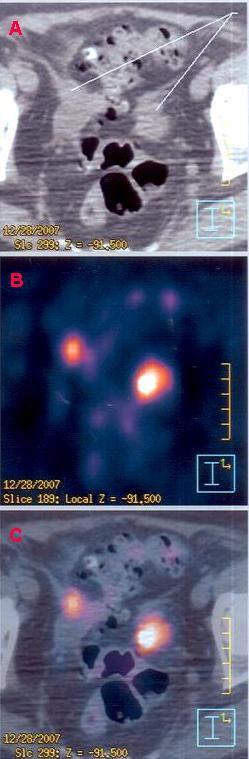
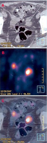
English: Excerpt of PET-CT clinical images from a 69-yrs old female with metastases in the pelvic area, evidenced by hypermetabolic spots, one near the sigmoid colon (at right), another in a lymphatic node (at left). Obtained with a Philips PET-CT device in the Cancer Hospital of São Paulo. Patient identification is not shown. |
| Source | http://en.wikipedia.org/wiki/Image:Pet-ct-images.jpg |
| Author | Renato M.E. Sabbatini, PhD |
Licensing edit
This file is licensed under the Creative Commons Attribution 3.0 Unported license.
- You are free:
- to share – to copy, distribute and transmit the work
- to remix – to adapt the work
- Under the following conditions:
- attribution – You must give appropriate credit, provide a link to the license, and indicate if changes were made. You may do so in any reasonable manner, but not in any way that suggests the licensor endorses you or your use.
File history
Click on a date/time to view the file as it appeared at that time.
| Date/Time | Thumbnail | Dimensions | User | Comment | |
|---|---|---|---|---|---|
| current | 09:33, 17 October 2008 | 249 × 759 (32 KB) | Formol (talk | contribs) | {{Information |Description={{en|1=Excerpt of PET-CT clinical images from a 69-yrs old female with metastases in the pelvic area, evidenced by hypermetabolic spots, one near the sigmoid colon (at right), another in a lymphatic node (at left). Obtained with |
You cannot overwrite this file.
File usage on Commons
The following page uses this file:
File usage on other wikis
The following other wikis use this file:
- Usage on cs.wikipedia.org
- Usage on en.wikipedia.org
- Usage on fa.wikipedia.org
- Usage on he.wikipedia.org
- Usage on ko.wikipedia.org
- Usage on pt.wikipedia.org