File:Posterior view of the virtual reconstruction of DH3.jpg
Size of this preview: 672 × 600 pixels. Other resolutions: 269 × 240 pixels | 538 × 480 pixels | 707 × 631 pixels.
Original file (707 × 631 pixels, file size: 68 KB, MIME type: image/jpeg)
Captions
Captions
Add a one-line explanation of what this file represents
Summary edit
| Description |
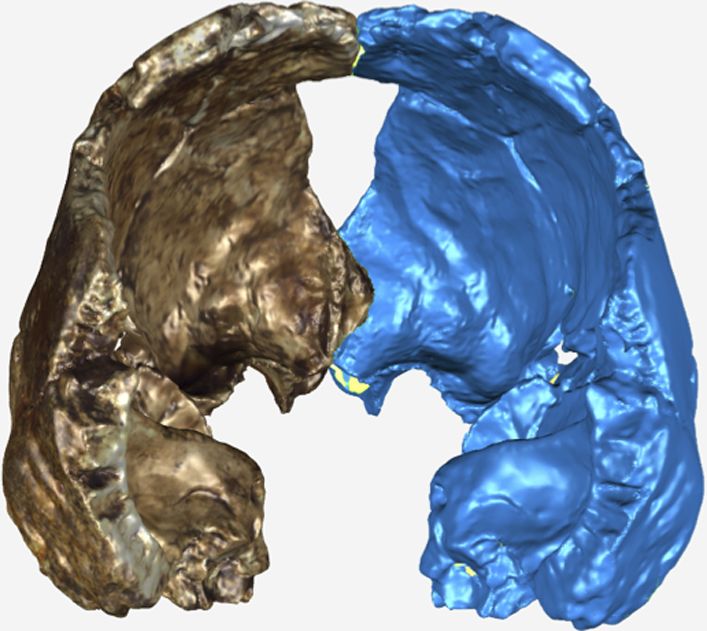
English: Posterior view of the virtual reconstruction of DH3. The resultant mirror image is displayed in blue. The antimeres were aligned by the frontal crest and sagittal suture using the Manual Registration function in GeoMagic Studio 14.0. |
| Date | |
| Source | http://elifesciences.org/content/4/e09560 |
| Author | Lee Roger Berger research team |
Licensing edit
This file is licensed under the Creative Commons Attribution-Share Alike 4.0 International license.
- You are free:
- to share – to copy, distribute and transmit the work
- to remix – to adapt the work
- Under the following conditions:
- attribution – You must give appropriate credit, provide a link to the license, and indicate if changes were made. You may do so in any reasonable manner, but not in any way that suggests the licensor endorses you or your use.
- share alike – If you remix, transform, or build upon the material, you must distribute your contributions under the same or compatible license as the original.
File history
Click on a date/time to view the file as it appeared at that time.
| Date/Time | Thumbnail | Dimensions | User | Comment | |
|---|---|---|---|---|---|
| current | 20:10, 17 September 2015 | | 707 × 631 (68 KB) | Mały koleżka (talk | contribs) | User created page with UploadWizard |
You cannot overwrite this file.
File usage on Commons
The following page uses this file: