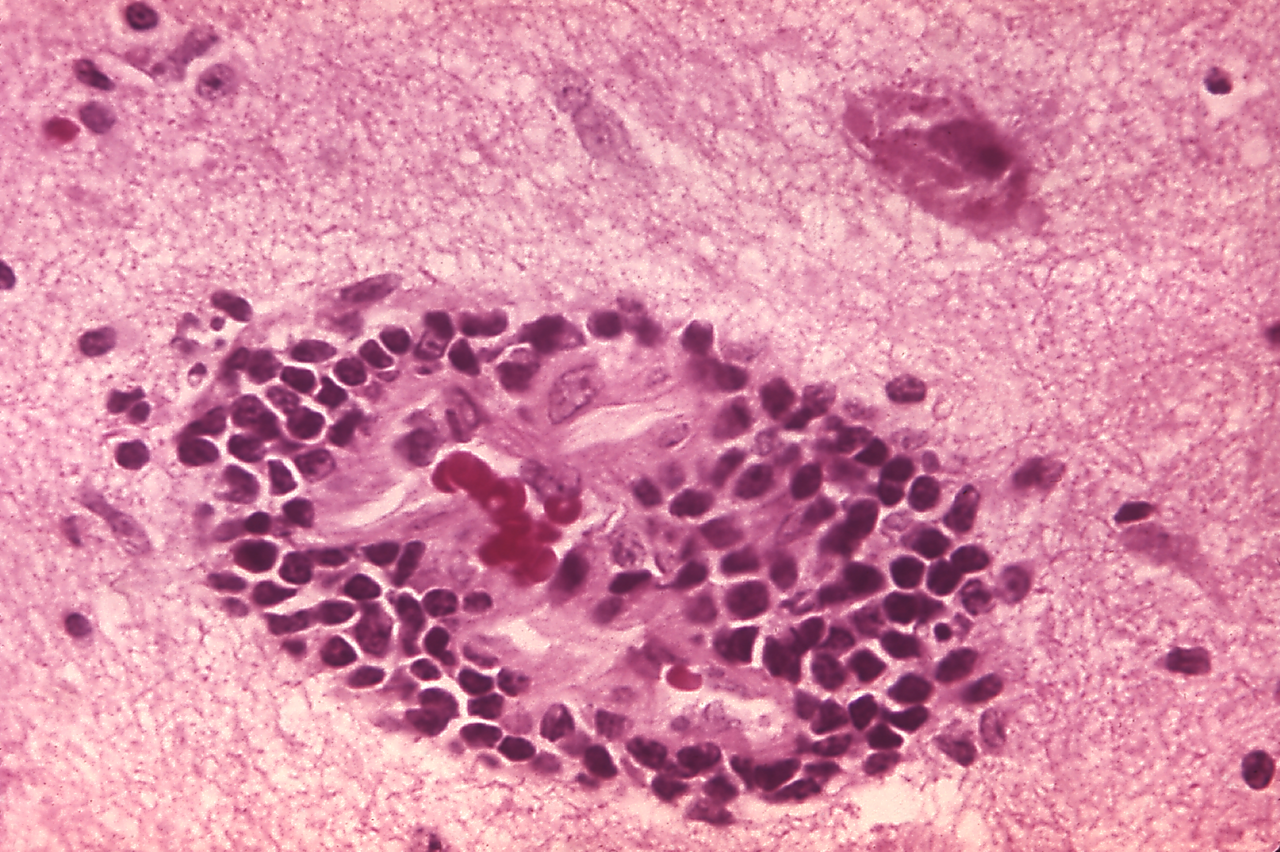
File:Rabies encephalitis PHIL 3368.png

Size of this preview: 800 × 533 pixels. Other resolutions: 320 × 213 pixels | 640 × 426 pixels | 1,024 × 682 pixels | 1,280 × 852 pixels | 1,801 × 1,199 pixels.
Original file (1,801 × 1,199 pixels, file size: 4.06 MB, MIME type: image/png)
Captions
Captions
Add a one-line explanation of what this file represents
Summary edit
| Description | |||
| Date | |||
| Source |
|
||
| Author | Content Provider(s): CDC/Dr. Daniel P. Perl | ||
| Permission (Reusing this file) |
Copyright Restrictions: None – This image is in the public domain and thus free of any copyright restrictions. As a matter of courtesy we request that the content provider be credited and notified in any public or private usage of this image. | ||
| Other versions | Rabies encephalitis PHIL 3368 lores.jpg (same resolution JPEG image). |
Licensing edit
This image is a work of the Centers for Disease Control and Prevention, part of the United States Department of Health and Human Services, taken or made as part of an employee's official duties. As a work of the U.S. federal government, the image is in the public domain.
|
File history
Click on a date/time to view the file as it appeared at that time.
| Date/Time | Thumbnail | Dimensions | User | Comment | |
|---|---|---|---|---|---|
| current | 07:52, 26 October 2011 | | 1,801 × 1,199 (4.06 MB) | Ghainmem (talk | contribs) |
You cannot overwrite this file.
File usage on Commons
There are no pages that use this file.