File:STD Depth Coded Stack Phallodin Stained Actin Filaments.png

Original file (4,096 × 4,096 pixels, file size: 21.54 MB, MIME type: image/png)
Captions
Captions
Summary edit
| Description |
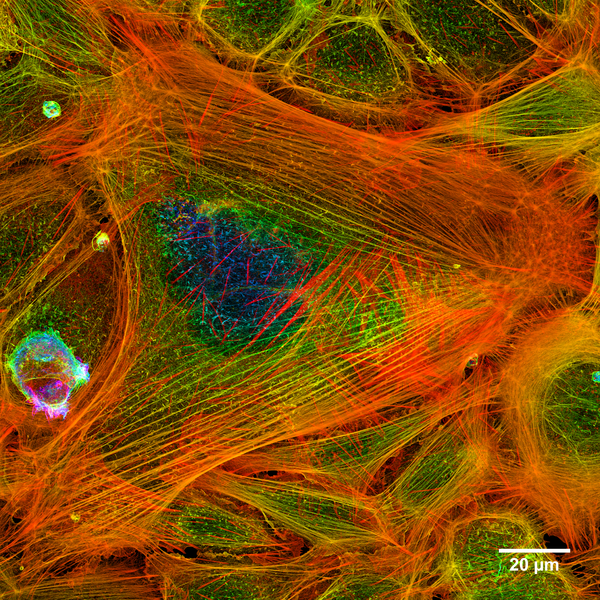
Čeština: Vizualizovaná aktinová mikrofilamenta, fluorescenčně značená faloidinem, podílející se na tvorbě buněčného cytoskeletu.
English: A merged stack of confocal images showing actin filaments within a cell. The image has been colour coded in the z axis to show in a 2D image which heights filaments can be found at within cells.
Dimensions Español: Un conjunto fusionado de imágenes confocales que muestran los filamentos de actina en una celula.
Українська: Суміщені конфокальні зображення мікрофіламентів актину в клітині. Висота мікрофіламентів кодована кольором.
Polski: Złożenie zdjęć wykonanych za pomocą mikroskopu konfokalnego przedstawiające filamenty aktyny wewnątrz komórki. W celu lepszego odwzorowania na płaszczyźnie, położenie filamentów względem osi Z zostało „zakodowane” za pomocą kolorów.
Português: Imagem construída por microscopia confocal mostrando filamentos de actina no interior de uma célula.
Bosanski: Spoj više konfokalnih slika prikazuje aktinske filamente unutar ćelije.
Bahasa Indonesia: Gambar sebuah gabungan pembuluh menunjukan jaringan filamen dengan sel.
Italiano: Una pila sovrapposta di immagini confocali che mostra dei filamenti di actina all'interno di una cellula.
한국어: 세포내 가는근육잔섬유의 공초점 이미지 중첩 합성.
|
| Date | |
| Source | Own work |
| Author | Howard Vindin |
Assessment edit
|

This image was selected as picture of the day on Wikimedia Commons for 24 July 2015. It was captioned as follows: English: A merged stack of confocal images showing actin filaments within a cell. Other languages:
Bahasa Indonesia: Gambar sebuah gabungan pembuluh menunjukan jaringan filamen dengan sel. Bosanski: Spoj više konfokalnih slika prikazuje aktinske filamente unutar ćelije. Čeština: Vizualizovaná aktinová mikrofilamenta, fluorescenčně značená faloidinem, podílející se na tvorbě buněčného cytoskeletu. English: A merged stack of confocal images showing actin filaments within a cell. Español: Un conjunto fusionado de imágenes confocales que muestran los filamentos de actina en una celula. Italiano: Una pila sovrapposta di immagini confocali che mostra dei filamenti di actina all'interno di una cellula. Português: Imagem construída por microscopia confocal mostrando filamentos de actina no interior de uma célula. Sunda: Gabungan urat saraf, ngambar mintonkeun ramat filamén jeung sél. Українська: Суміщені конфокальні зображення мікрофіламентів актину в клітині. Висота мікрофіламентів кодована кольором 한국어: 세포내 가는근육잔섬유의 공초점 이미지 중첩 합성. |
This image was selected as picture of the day on Vietnamese Wikipedia.
|
Licensing edit
- You are free:
- to share – to copy, distribute and transmit the work
- to remix – to adapt the work
- Under the following conditions:
- attribution – You must give appropriate credit, provide a link to the license, and indicate if changes were made. You may do so in any reasonable manner, but not in any way that suggests the licensor endorses you or your use.
- share alike – If you remix, transform, or build upon the material, you must distribute your contributions under the same or compatible license as the original.
File history
Click on a date/time to view the file as it appeared at that time.
| Date/Time | Thumbnail | Dimensions | User | Comment | |
|---|---|---|---|---|---|
| current | 00:33, 20 July 2015 | | 4,096 × 4,096 (21.54 MB) | Cmdrjameson (talk | contribs) | Compressed with pngout. Reduced by 9833kB (30% decrease). |
| 10:02, 24 April 2015 |  | 4,096 × 4,096 (31.14 MB) | Methylated603 (talk | contribs) | Added Scalebar | |
| 23:17, 6 April 2015 |  | 4,096 × 4,096 (27.34 MB) | Methylated603 (talk | contribs) | User created page with UploadWizard |
You cannot overwrite this file.
File usage on Commons
The following 36 pages use this file:
- User:Jtneill/Gallery
- User:Mattes/Favorite files/Images/B
- User:Methylated603
- User talk:Methylated603
- Commons:Featured picture candidates/File:STD Depth Coded Stack Phallodin Stained Actin Filaments.png
- Commons:Featured picture candidates/Log/April 2015
- Commons:Featured pictures/Natural phenomena
- Commons:Featured pictures/chronological/2015-A
- Commons:Picture of the Year/2015/Candidates
- Commons:Picture of the Year/2015/Candidates/R2
- Commons:Picture of the Year/2015/R1/Gallery/2015-A
- Commons:Picture of the Year/2015/R1/Gallery/ALL
- Commons:Picture of the Year/2015/R1/Gallery/M04
- Commons:Picture of the Year/2015/R1/Gallery/Maps
- Commons:Picture of the Year/2015/R1/Results/Finalists
- Commons:Picture of the Year/2015/R1/Results/Top (non strict)
- Commons:Picture of the Year/2015/R1/Results/Top (strict)
- Commons:Picture of the Year/2015/R1/v/STD Depth Coded Stack Phallodin Stained Actin Filaments.png
- Commons:Picture of the Year/2015/R2/Autoresults
- Commons:Picture of the Year/2015/R2/Gallery
- Commons:Picture of the Year/2015/R2/Results/Candidates/Gallery
- Commons:Picture of the Year/2015/R2/v/STD Depth Coded Stack Phallodin Stained Actin Filaments.png
- Template:Potd/2015-07
- Template:Potd/2015-07-24
- Template:Potd/2015-07-24 (bs)
- Template:Potd/2015-07-24 (cs)
- Template:Potd/2015-07-24 (en)
- Template:Potd/2015-07-24 (es)
- Template:Potd/2015-07-24 (hu)
- Template:Potd/2015-07-24 (id)
- Template:Potd/2015-07-24 (it)
- Template:Potd/2015-07-24 (ko)
- Template:Potd/2015-07-24 (nl)
- Template:Potd/2015-07-24 (pt)
- Template:Potd/2015-07-24 (su)
- Template:Potd/2015-07-24 (uk)
File usage on other wikis
The following other wikis use this file:
- Usage on ar.wikipedia.org
- Usage on be-tarask.wikipedia.org
- Usage on bn.wikipedia.org
- Usage on ca.wikipedia.org
- Usage on crh.wikipedia.org
- Usage on cs.wikipedia.org
- Usage on cv.wikipedia.org
- Usage on de.wikipedia.org
- Usage on en.wikipedia.org
- Usage on es.wikipedia.org
- Usage on eu.wikipedia.org
- Usage on fr.wikipedia.org
- Usage on he.wikipedia.org
- Usage on hu.wikipedia.org
- Usage on id.wikipedia.org
- Usage on it.wiktionary.org
- Usage on ka.wikipedia.org
- Usage on ko.wikipedia.org
- Usage on la.wikipedia.org
- Usage on lbe.wikipedia.org
- Usage on mk.wikipedia.org
- Usage on nl.wikipedia.org
- Usage on os.wikipedia.org
- Usage on pt.wikipedia.org
- Usage on ru.wikipedia.org
- Usage on ru.wikinews.org
- Usage on sah.wikipedia.org
- Usage on su.wikipedia.org
- Usage on tr.wikipedia.org
- Usage on tt.wikipedia.org
- Usage on uk.wikipedia.org
- Usage on vi.wikipedia.org
View more global usage of this file.