File:Schleiden; cellular tissue of plants Wellcome M0010608.jpg

Original file (2,550 × 4,159 pixels, file size: 4 MB, MIME type: image/jpeg)
Captions
Captions
Summary edit
| Schleiden; cellular tissue of plants | |||
|---|---|---|---|
| Title |
Schleiden; cellular tissue of plants |
||
| Description |
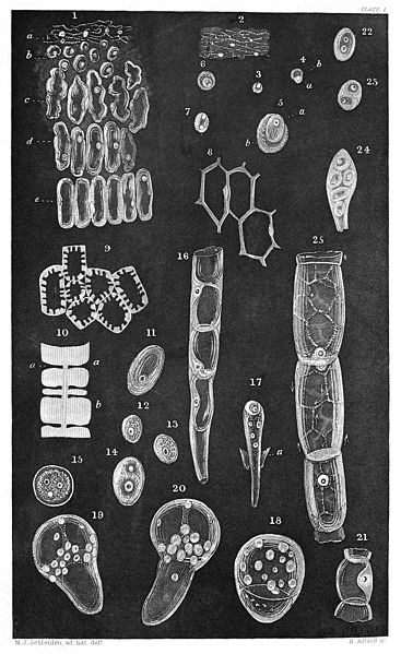
Appended to Schwann's book in the same volume; Contributions to Phytogenesis, translated from the German of Dr M. J. Schleiden, Professor of Botany at the University of Jena. Plate I Fig. 1. Cellular tissue from the embryo-sac of Chamoedorea Scheideana in the act of formation. a. The inner-most mass, consisting of bum with intermingled mucous granules and cytoblasts. b. Newly formed cells, still soluble in distilled water. c-e. Further development of the cells, which, with the exception of the cytoblasts, may still coalesce, under slight pressure, into an amorphous mass. 2. the formative substance from fig. 1, a, more highly magnified, bum, mucous granules, nuclei of the cytoblasts, and cytoblasts. 3. a single and as yet free cytoblast, still more highly magnified. 4. A cytoblast with the cell forming upon it. 5. The same, more highly magnified. 6. The same. The cytoblast here exhibits two nuclei, and is delineated in 7, isolated after the destruction of the cell by pressure. 8. the same cellular tissue in a higher degree of development that that represented by fig. 1, e. The contiguous cell-walls have already united. By making a transverse section, it may be distinctly perceived the that cytoblast is enclosed in the cell-wall. 9. Cells from a delicate transverse sectionof the almost matured albumen. 10. Common partition-wall between two cells from fig. 9, under a higher magnifying power. The stratiform depositions may be ovserved at b, and the porous canals produced by their local failure at a. I could distinctly enumerate from nine to twelve layers which had been deposited within fourteen days. 11. a sporule from Rhizina laevigata Fries, with the cytoblast. 12, 13, 14. Different cytoblasts from the embryo-sac of Pimelea drupacea before the appearance of cells. 15. A young cell with its cytoblast, from the same. The latter in this instance presents the unusual number of three nucleoli. 16. A portion of the embryonal end of the pollen-tube projecting from the ovulum in Orchis Morio, within which, towards the upper part, cells have been already developed. At the lower part, the original pollen-tube may still be distinguished. The almost globular cytoblasts are, in this instance, distinctly enclosed in the cell-wall. 17. Embryonal end of the pollen-tube from Linum pallescens, together with an appended lobule of the embryo-sac (a). The process of the formation of cells is commencing. Above, a young cell with its cytoblast is already perceptible, beneath this several cell-nuclei are seen floating free. 18, 19, 20. Commencing germination in the sporules of Marchantia polymorpha. Compare the text, p. 248. 21. Portions of the pollen-tube which have become cellular, from Orchis latifolia, in the highest stage of development; the investment of the pollen-tube is no longer perceptible. the cytoblast is enclosed in the cell-wall, just as in fig. 16. 22 and 23. Two isolated cells from the terminal shoot (punctum vegetationis, Wolff) of Gasteria racemosa; 22 exhibitis two free cytoblasts; 23, two newly-formed cells within the original cell. Fig 24. A very young leaf of Crassula portulaca, the five cells which solely compose it being still surrounded by a parent cell. 25. Three cells from an articulated hair of potato, with a retiform current of mucus upon their walls. In the central cell the direction of the currents is partially indicated by arrows. Rare Books |
||
| Credit line |
|
||
| References |
|
||
| Source/Photographer |
https://wellcomeimages.org/indexplus/obf_images/55/5c/5207ca96c667b2f07440c28cd4b1.jpg
|
||
| Other versions |
  |
||
Licensing edit
- You are free:
- to share – to copy, distribute and transmit the work
- to remix – to adapt the work
- Under the following conditions:
- attribution – You must give appropriate credit, provide a link to the license, and indicate if changes were made. You may do so in any reasonable manner, but not in any way that suggests the licensor endorses you or your use.
File history
Click on a date/time to view the file as it appeared at that time.
| Date/Time | Thumbnail | Dimensions | User | Comment | |
|---|---|---|---|---|---|
| current | 21:18, 23 October 2014 |  | 2,550 × 4,159 (4 MB) | Fæ (talk | contribs) | =={{int:filedesc}}== {{Artwork |artist = |author = |title = Schleiden; cellular tissue of plants |description = Appended to Schwann's book in the same volume; Contributions to Phytogenesis, translated from th... |
You cannot overwrite this file.
File usage on Commons
The following 3 pages use this file:
Metadata
This file contains additional information such as Exif metadata which may have been added by the digital camera, scanner, or software program used to create or digitize it. If the file has been modified from its original state, some details such as the timestamp may not fully reflect those of the original file. The timestamp is only as accurate as the clock in the camera, and it may be completely wrong.
| Short title | M0010608 Schleiden; cellular tissue of plants |
|---|---|
| Author | Wellcome Library, London |
| Headline | M0010608 Schleiden; cellular tissue of plants |
| Copyright holder | Copyrighted work available under Creative Commons Attribution only licence CC BY 4.0 http://creativecommons.org/licenses/by/4.0/ |
| Image title | M0010608 Schleiden; cellular tissue of plants
Credit: Wellcome Library, London. Wellcome Images images@wellcome.ac.uk http://wellcomeimages.org Appended to Schwann's book in the same volume; Contributions to Phytogenesis, translated from the German of Dr M. J. Schleiden, Professor of Botany at the University of Jena. Plate I Fig. 1. Cellular tissue from the embryo-sac of Chamoedorea Scheideana in the act of formation. a. The inner-most mass, consisting of bum with intermingled mucous granules and cytoblasts. b. Newly formed cells, still soluble in distilled water. c-e. Further development of the cells, which, with the exception of the cytoblasts, may still coalesce, under slight pressure, into an amorphous mass. 2. the formative substance from fig. 1, a, more highly magnified, bum, mucous granules, nuclei of the cytoblasts, and cytoblasts. 3. a single and as yet free cytoblast, still more highly magnified. 4. A cytoblast with the cell forming upon it. 5. The same, more highly magnified. 6. The same. The cytoblast here exhibits two nuclei, and is delineated in 7, isolated after the destruction of the cell by pressure. 8. the same cellular tissue in a higher degree of development that that represented by fig. 1, e. The contiguous cell-walls have already united. By making a transverse section, it may be distinctly perceived the that cytoblast is enclosed in the cell-wall. 9. Cells from a delicate transverse sectionof the almost matured albumen. 10. Common partition-wall between two cells from fig. 9, under a higher magnifying power. The stratiform depositions may be ovserved at b, and the porous canals produced by their local failure at a. I could distinctly enumerate from nine to twelve layers which had been deposited within fourteen days. 11. a sporule from Rhizina laevigata Fries, with the cytoblast. 12, 13, 14. Different cytoblasts from the embryo-sac of Pimelea drupacea before the appearance of cells. 15. A young cell with its cytoblast, from the same. The latte |
| IIM version | 2 |
