File:Serum protein electrophoresis normal and paraprotein.svg

Size of this PNG preview of this SVG file: 440 × 599 pixels. Other resolutions: 176 × 240 pixels | 352 × 480 pixels | 564 × 768 pixels | 752 × 1,024 pixels | 1,504 × 2,048 pixels | 492 × 670 pixels.
Original file (SVG file, nominally 492 × 670 pixels, file size: 30 KB)
Captions
Captions
Add a one-line explanation of what this file represents
Summary
edit| Description |
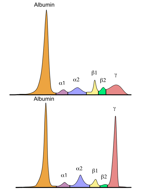
English: An example serum protein electrophoresis. Albumin (orange), alpha-1 (pink), alpha-2 (blue), beta-1 (yellow), beta-2 (green) and gamma (red) peaks are shown. Antibodies are mostly found in the gamma region. The top image shows serum protein electrophoresis from a normal individual with polyclonal antibodies (no peaks in the gamma region). The bottom image a monoclonal antibody present (paraprotein), which presents as a peak in the gamma region.
Deutsch: Serumeiweißelektrophorese mit den verschiedenen Proteinfraktionen. Oben: Normalbefund, unten: monoklonale Vermehrung von Immunglobulinen (Paraproteinämie). |
|||
| Date | ||||
| Source | Serum_protein_electrophoresis_normal_and_paraprotein.png by Simon Caulton | |||
| Author | Furfur | |||
| Permission (Reusing this file) |
I, the copyright holder of this work, hereby publish it under the following licenses:
This file is licensed under the Creative Commons Attribution-Share Alike 4.0 International license.
You may select the license of your choice. |
|||
| Other versions |
|
|||
| SVG development | This vector image was created with Adobe Illustrator. |


File history
Click on a date/time to view the file as it appeared at that time.
| Date/Time | Thumbnail | Dimensions | User | Comment | |
|---|---|---|---|---|---|
| current | 20:27, 5 June 2016 | | 492 × 670 (30 KB) | Furfur (talk | contribs) | Detail |
| 20:23, 5 June 2016 |  | 492 × 670 (29 KB) | Furfur (talk | contribs) | =={{int:filedesc}}== {{Information |description={{en|1=An example serum protein electrophoresis. Albumin (orange), alpha-1 (pink), alpha-2 (blue), beta-1 (yellow), beta-2 (green) and gamma (red) peaks are shown. Antibodies are mostly found in the gamm... |
You cannot overwrite this file.
File usage on Commons
The following page uses this file:
File usage on other wikis
The following other wikis use this file:
- Usage on de.wikipedia.org
- Usage on ja.wikipedia.org
- Usage on no.wikipedia.org