File:Shiga toxin 1DM0.png

Size of this preview: 747 × 600 pixels. Other resolutions: 299 × 240 pixels | 598 × 480 pixels | 956 × 768 pixels | 1,275 × 1,024 pixels | 1,848 × 1,484 pixels.
Original file (1,848 × 1,484 pixels, file size: 1.06 MB, MIME type: image/png)
Captions
Captions
Add a one-line explanation of what this file represents
| Description |
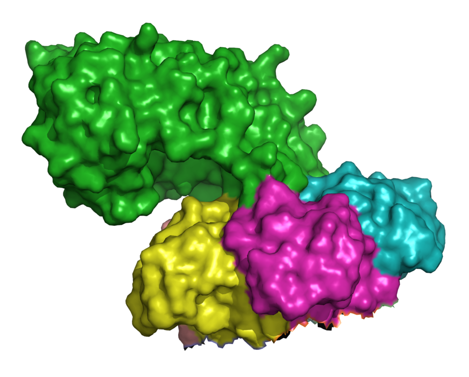
English: 3d surface model of shiga toxin A+5B subunits (stxB from Shigella dyasenteriae) from PDB 1DMO. Ref.: M.E.Fraser et al. (1994). Crystal structure of the holotoxin from Shigella dysenteriae at 2.5 A resolution.. Nat Struct Biol, 1, 59-64. PMID 7656009 [DOI: 10.1038/nsb0194-59] |
||
| Date | |||
| Source | adapted from http://www.pdb.org/pdb/files/1dm0.pdb using PyMOL | ||
| Author | own work | ||
| Permission (Reusing this file) |
|
||
| Other versions | http://www.ebi.ac.uk/pdbsum/1DM0 |
File history
Click on a date/time to view the file as it appeared at that time.
| Date/Time | Thumbnail | Dimensions | User | Comment | |
|---|---|---|---|---|---|
| current | 09:10, 27 October 2008 | | 1,848 × 1,484 (1.06 MB) | Ayacop (talk | contribs) | {{Information |Description={{en|1=3d ribbon model of shiga toxin A+5B subunits (stxB from ''Shigella dyasenteriae'') from PDB 1DMO. Ref.: M.E.Fraser et al. (1994). Crystal structure of the holotoxin from Shigella dysenteriae at 2.5 A resolution.. Nat Stru |
You cannot overwrite this file.
File usage on Commons
There are no pages that use this file.
File usage on other wikis
The following other wikis use this file:
- Usage on de.wikipedia.org
- Usage on fr.wikipedia.org
- Usage on he.wikipedia.org