File:Telocytes - Fig 2.jpg

Size of this preview: 800 × 458 pixels. Other resolutions: 320 × 183 pixels | 640 × 366 pixels | 1,024 × 586 pixels | 1,280 × 732 pixels | 1,947 × 1,114 pixels.
Original file (1,947 × 1,114 pixels, file size: 2 MB, MIME type: image/jpeg)
Captions
Captions
Add a one-line explanation of what this file represents
Summary
edit
| Description |
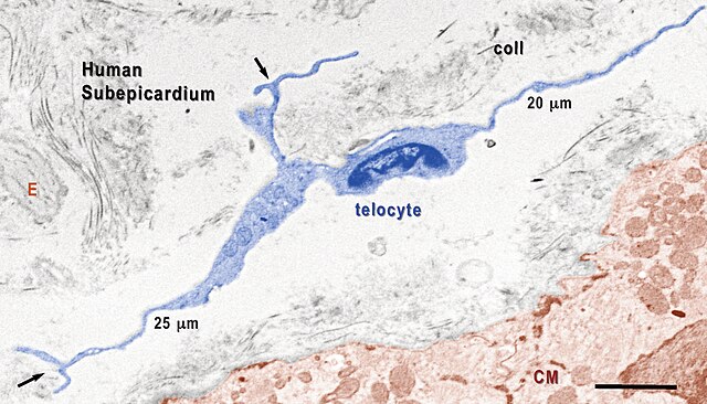
English: Digitally coloured TEM image shows TC (blue) in human subepicardium, bordering the peripheral cardiomyocytes (CM, highlighted in brown). The TC has three telopodes, illustrating: a) the distinctive dichotomous pattern of branching (arrows); b) Tp are very thin at the emergence of the cell body; c) alternating podoms and podomeres. Note that some portions of podomeres have the same thickness as collagen fibrills, which make them impossible to be observed under light microscopy. E - elastin Scale bar - 2 mm. |
| Date | |
| Source | Own work |
| Author | Lmpopescu |
Licensing
edit
I, the copyright holder of this work, hereby publish it under the following licenses:
This file is licensed under the Creative Commons Attribution-Share Alike 3.0 Unported license.
- You are free:
- to share – to copy, distribute and transmit the work
- to remix – to adapt the work
- Under the following conditions:
- attribution – You must give appropriate credit, provide a link to the license, and indicate if changes were made. You may do so in any reasonable manner, but not in any way that suggests the licensor endorses you or your use.
- share alike – If you remix, transform, or build upon the material, you must distribute your contributions under the same or compatible license as the original.

|
Permission is granted to copy, distribute and/or modify this document under the terms of the GNU Free Documentation License, Version 1.2 or any later version published by the Free Software Foundation; with no Invariant Sections, no Front-Cover Texts, and no Back-Cover Texts. A copy of the license is included in the section entitled GNU Free Documentation License. |
You may select the license of your choice.
File history
Click on a date/time to view the file as it appeared at that time.
| Date/Time | Thumbnail | Dimensions | User | Comment | |
|---|---|---|---|---|---|
| current | 12:24, 18 March 2011 | | 1,947 × 1,114 (2 MB) | Lmpopescu (talk | contribs) | {{Information |Description ={{en|1=Digitally coloured TEM image shows TC (blue) in human subepicardium, bordering the peripheral cardiomyocytes (CM, highlighted in brown). The TC has three telopodes, illustrating: a) the distinctive dichotomous pattern |
You cannot overwrite this file.
File usage on Commons
There are no pages that use this file.
File usage on other wikis
The following other wikis use this file: