File:Tissue-resident macrophages in the developing testes.jpg

Original file (2,119 × 1,372 pixels, file size: 2.4 MB, MIME type: image/jpeg)
Captions
Captions
Summary
edit
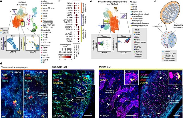
| Description | Figure 5. Tissue-resident macrophages in the developing testes. Transcriptional, spatiotemporal and paracrine signatures of human pregranulosa cells.a, UMAP of immune cell states (colour) in the human scRNA-seq data (n = 20,556). Doublets and low-quality control cells were removed. Eleven samples were enriched for immune (CD45+) cells. Zoomed-in UMAPs show SIGLEC15+ and TREM2+ fetal testicular macrophages (ftMs) labelled by sex. b, Dot plot showing variance-scaled, log-transformed expression of marker genes (y-axis) for the identified macrophage subsets (x-axis). c, UMAP projections of integrated myeloid cells (colour) from several embryonic/fetal tissues (n = 58,948). Zoomed-in UMAPs show osteoclast and microglia signature macrophages labelled by tissue of origin. d, High-resolution imaging of representative human gonadal sections with intensity proportional to smFISH signal for RNA markers. Left, 12 PCW testis and ovary stained for CD68 (yellow, macrophages), F13A1 (red, tissue-repair macrophages) and NR2F2 (cyan, mesenchymal) (n = 2). Middle, 12 PCW testis stained for PDGFRA (green, mesenchymal), CDH5 (cyan, endothelial), CD68 (red, macrophages) and SIGLEC15 (yellow, SIGLEC15+ ftMs). SIGLEC15+ ftMs (white arrows) are outside the testis cords in proximity to endothelial cells (n = 5). Right, 8 PCW testis stained for SOX9 (magenta, Sertoli (n = 5)), POU5F1 (magenta, PGCs (n = 2)), CD68 (red, macrophages), P2RY12 (yellow, TREM2+ ftMs) and PDGFRA (cyan, mesenchymal). TREM2+ ftMs (white arrows) are adjacent to the germ and Sertoli cells. White dashed rectangles highlight gonadal regions magnified; scale bars, 100 and 10 µm in magnified regions; testicular developing cords are delineated with dashed lines. e, Schematics illustrating the spatial location of the distinct testicular macrophage populations. cDC, conventional dendritic cells; ftM, fetal testicular macrophages; ILC, innate lymphoid cells; mega, megakaryocytes; MEMP, megakaryocyte-erythroid-mast cell progenitors; mono, monocytes; neutro, neutrophils; NMP, neutrophil-myeloid progenitors; NK, natural killer cells; pDC, plasmacytoid dendritic cell; prec, precursor; Pre-B, pre-B cells; Pre-pro-B, pre-pro-B cells; Pro-B, pro-B cells; prog, progenitor; T, T cells. |
| Date | |
| Source | https://www.nature.com/articles/s41586-022-04918-4/ Single-cell roadmap of human gonadal development. Nature 607, 540–547 (2022). https://doi.org/10.1038/s41586-022-04918-4 |
| Author | Garcia-Alonso, L., Lorenzi, V., Mazzeo, C.I. et al. |

|
This file, which was originally posted to an external website, has not yet been reviewed by an administrator or reviewer to confirm that the above license is valid. See Category:License review needed for further instructions.
|
Open Access This article is licensed under a Creative Commons Attribution 4.0 International License, which permits use, sharing, adaptation, distribution and reproduction in any medium or format, as long as you give appropriate credit to the original author(s) and the source, provide a link to the Creative Commons license, and indicate if changes were made. The images or other third party material in this article are included in the article’s Creative Commons license, unless indicated otherwise in a credit line to the material. If material is not included in the article’s Creative Commons license and your intended use is not permitted by statutory regulation or exceeds the permitted use, you will need to obtain permission directly from the copyright holder. To view a copy of this license, visit https://creativecommons.org/licenses/by/4.0/.
Licensing
edit
- You are free:
- to share – to copy, distribute and transmit the work
- to remix – to adapt the work
- Under the following conditions:
- attribution – You must give appropriate credit, provide a link to the license, and indicate if changes were made. You may do so in any reasonable manner, but not in any way that suggests the licensor endorses you or your use.
File history
Click on a date/time to view the file as it appeared at that time.
| Date/Time | Thumbnail | Dimensions | User | Comment | |
|---|---|---|---|---|---|
| current | 17:21, 10 July 2024 | | 2,119 × 1,372 (2.4 MB) | Rasbak (talk | contribs) | {{Information |description=Figure 5. Tissue-resident macrophages in the developing testes. Transcriptional, spatiotemporal and paracrine signatures of human pregranulosa cells.a, UMAP of immune cell states (colour) in the human scRNA-seq data (n = 20,556). Doublets and low-quality control cells were removed. Eleven samples were enriched for immune (CD45+) cells. Zoomed-in UMAPs show SIGLEC15+ and TREM2+ fetal testicular macrophages (ftMs) labelled by sex. b, Dot plot showing variance-scaled,... |
You cannot overwrite this file.
File usage on Commons
There are no pages that use this file.