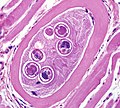
File:Trichinella BAM5b.jpg
Trichinella_BAM5b.jpg (300 × 300 pixels, file size: 75 KB, MIME type: image/jpeg)
File information
Structured data
Captions
Captions
Add a one-line explanation of what this file represents
Summary
edit| DescriptionTrichinella BAM5b.jpg |
English: Figure E: Higher-magnification of the larvae in Figure C. Shown in these cuts are a nucleated stichocyte (ST), prominent lateral chords, or bacillary bands, (LC), immature reproductive tubes (RT), and the intestine (IN). Image captured at 1000x magnification.
|
| Date | |
| Source | https://www.cdc.gov/dpdx/trichinellosis/gallery.html#encysted |
| Author | CDC DPDx |
Licensing
edit| Public domainPublic domainfalsefalse |
This image is a work of the Centers for Disease Control and Prevention, part of the United States Department of Health and Human Services, taken or made as part of an employee's official duties. As a work of the U.S. federal government, the image is in the public domain.
|
 |
File history
Click on a date/time to view the file as it appeared at that time.
| Date/Time | Thumbnail | Dimensions | User | Comment | |
|---|---|---|---|---|---|
| current | 23:41, 25 July 2015 |  | 300 × 300 (75 KB) | CFCF (talk | contribs) | User created page with UploadWizard |
You cannot overwrite this file.
File usage on Commons
There are no pages that use this file.
Metadata
This file contains additional information such as Exif metadata which may have been added by the digital camera, scanner, or software program used to create or digitize it. If the file has been modified from its original state, some details such as the timestamp may not fully reflect those of the original file. The timestamp is only as accurate as the clock in the camera, and it may be completely wrong.
| Camera manufacturer | OLYMPUS |
|---|---|
| Camera model | DP72 |
| Author | gqa4 |
| Exposure time | 263/250,000 sec (0.001052) |
| Orientation | Normal |
| Horizontal resolution | 300 dpi |
| Vertical resolution | 300 dpi |
| Software used | Adobe Photoshop 7.0 |
| File change date and time | 10:54, 4 March 2011 |
| Exif version | 2.1 |
| Date and time of digitizing | 15:12, 4 March 2011 |
| APEX aperture | 0.4 |
| Subject distance | 0.003 meters |
| Metering mode | Spot |
| Supported Flashpix version | 1 |
| Color space | Uncalibrated |
| Exposure mode | Manual exposure |
| GPS tag version | 51.51.53.53.52.52.51.50 |


