File:Two-color MINFLUX image of mitochondria.jpg
Size of this preview: 785 × 600 pixels. Other resolutions: 314 × 240 pixels | 628 × 480 pixels | 827 × 632 pixels.
Original file (827 × 632 pixels, file size: 98 KB, MIME type: image/jpeg)
Captions
Captions
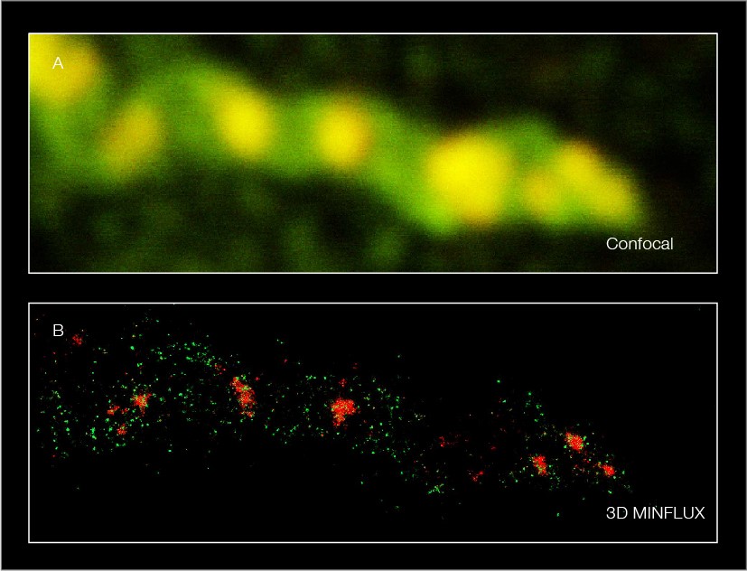
Comparison of mitochondria imaged with a confocal and a MINFLUX microscope.
Summary
edit
| Description |
English: The mitochondrial protein TOM20 (green) and mitochondrial DNA (red) were labeled in mammalian cells with indirect immunofluorescence using secondary antibodies coupled to sCy5 and CF680. Two-color confocal (A) and MINFLUX (B) imaging was performed using a ratiometric detection strategy. The labeling density of the two structures differs. For TOM20, single proteins are labeled in the mitochondrial membrane, whereas numerous binding sites are labeled in the mitochondrial DNA. MINFLUX enables visualizing and discriminating between the two structures. Scale bars: 500 nm. |
| Date | |
| Source | Images were provided by abberior Instruments GmbH |
| Author | abberior Instruments GmbH, contact person: Dr. Frederik Köpper |
| Permission (Reusing this file) |
Licensing
edit
This file is licensed under the Creative Commons Attribution-Share Alike 4.0 International license.
- You are free:
- to share – to copy, distribute and transmit the work
- to remix – to adapt the work
- Under the following conditions:
- attribution – You must give appropriate credit, provide a link to the license, and indicate if changes were made. You may do so in any reasonable manner, but not in any way that suggests the licensor endorses you or your use.
- share alike – If you remix, transform, or build upon the material, you must distribute your contributions under the same or compatible license as the original.
File history
Click on a date/time to view the file as it appeared at that time.
| Date/Time | Thumbnail | Dimensions | User | Comment | |
|---|---|---|---|---|---|
| current | 11:01, 11 August 2023 | | 827 × 632 (98 KB) | AByolia (talk | contribs) | Uploaded a work by abberior Instruments GmbH, contact person: Dr. Frederik Köpper from {{subst:PP}} Images were provided by abberior Instruments GmbH with UploadWizard |
You cannot overwrite this file.
File usage on Commons
There are no pages that use this file.