File:Unterschied Spinalanästhesie-Periduralanästhesie.png

Original file (1,200 × 531 pixels, file size: 187 KB, MIME type: image/png)
Captions
Captions
Summary edit
| DescriptionUnterschied Spinalanästhesie-Periduralanästhesie.png |
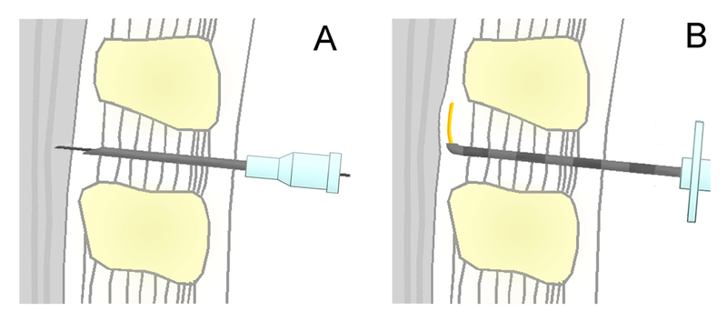
schematische Darstellung der Spinalanästhesie (A) und Periduralanästhesie (B) im Vergleich. Gezeigt ist ein medianer Schnitt in der Sagittalebene (Höhe mittlere Lendenwirbelsäule). Vereinfachte Darstellung, die Größenverhältnisse stimmen nicht mit der Realität überein. A. Prinzip der Durchführung einer Spinalanästhesie. Durch die zuvor zwischen den Dornfortsätzen der Wirbel L3/4 eingebrachte Führungskanüle wird die eigentliche Spinalnadel eingeführt und mit ihr der Subarachnoidalraum (Liquorraum) punktiert. Durch diese Nadel können dann Lokalanästhetika und andere Medikamente injiziert werden. B. Bei der Periduralanästhesie (PDA) wird eine Tuohy-Nadel vorgeschoben, bis der Periduralraum (Epiduralraum) erreicht wird, was sich durch einen Widerstandsverlust (Loss of Resistance) beim Einspritzen von Kochsalzlösung zeigt. durch die Kanüle wird dann der Periduralkatheter (gelb) in den Periduralraum eingeführt. Der Liquorraum ist bei einer PDA nicht betroffen. Inhaltliche Referenz: Jankovic, D: Regionalblockaden und Infiltrationstherapie. 3. Aufl. 2003. ABW Wissenschaftsverlagsgesellschaft. ISBN-13 9783936072167 |
| Date |
Spring 2008 date QS:P,+2008-00-00T00:00:00Z/9,P4241,Q40720559 |
| Source | Own work |
| Author | s. u. |
| Permission (Reusing this file) |
GFDL/CC-by-SA |
Licensing edit

|
Permission is granted to copy, distribute and/or modify this document under the terms of the GNU Free Documentation License, Version 1.2 or any later version published by the Free Software Foundation; with no Invariant Sections, no Front-Cover Texts, and no Back-Cover Texts. A copy of the license is included in the section entitled GNU Free Documentation License.http://www.gnu.org/copyleft/fdl.htmlGFDLGNU Free Documentation Licensetruetrue |
- You are free:
- to share – to copy, distribute and transmit the work
- to remix – to adapt the work
- Under the following conditions:
- attribution – You must give appropriate credit, provide a link to the license, and indicate if changes were made. You may do so in any reasonable manner, but not in any way that suggests the licensor endorses you or your use.
- share alike – If you remix, transform, or build upon the material, you must distribute your contributions under the same or compatible license as the original.
File history
Click on a date/time to view the file as it appeared at that time.
| Date/Time | Thumbnail | Dimensions | User | Comment | |
|---|---|---|---|---|---|
| current | 11:01, 21 March 2008 |  | 1,200 × 531 (187 KB) | PhilippN (talk | contribs) | {{Information |Description= |Source= |Date= |Author= |Permission= |other_versions= }} |
| 10:57, 21 March 2008 |  | 2,000 × 884 (364 KB) | PhilippN (talk | contribs) | == Beschreibung == {{Information |Description= schematische Darstellung der Spinalanästhesie (A) und Periduralanästhesie (B) im Vergleich. A. Prinzip der Durchführung einer [[:de:Spinalanästhesie |
You cannot overwrite this file.
File usage on Commons
There are no pages that use this file.
File usage on other wikis
The following other wikis use this file:
- Usage on de.wikipedia.org
- Usage on ja.wikipedia.org
- Usage on ru.wikipedia.org
- Usage on sr.wikipedia.org
- Usage on uz.wikipedia.org