File:West Nile Virus particles.jpg
Size of this preview: 604 × 599 pixels. Other resolutions: 242 × 240 pixels | 484 × 480 pixels | 774 × 768 pixels | 1,032 × 1,024 pixels | 1,792 × 1,778 pixels.
Original file (1,792 × 1,778 pixels, file size: 4.14 MB, MIME type: image/jpeg)
Captions
Captions
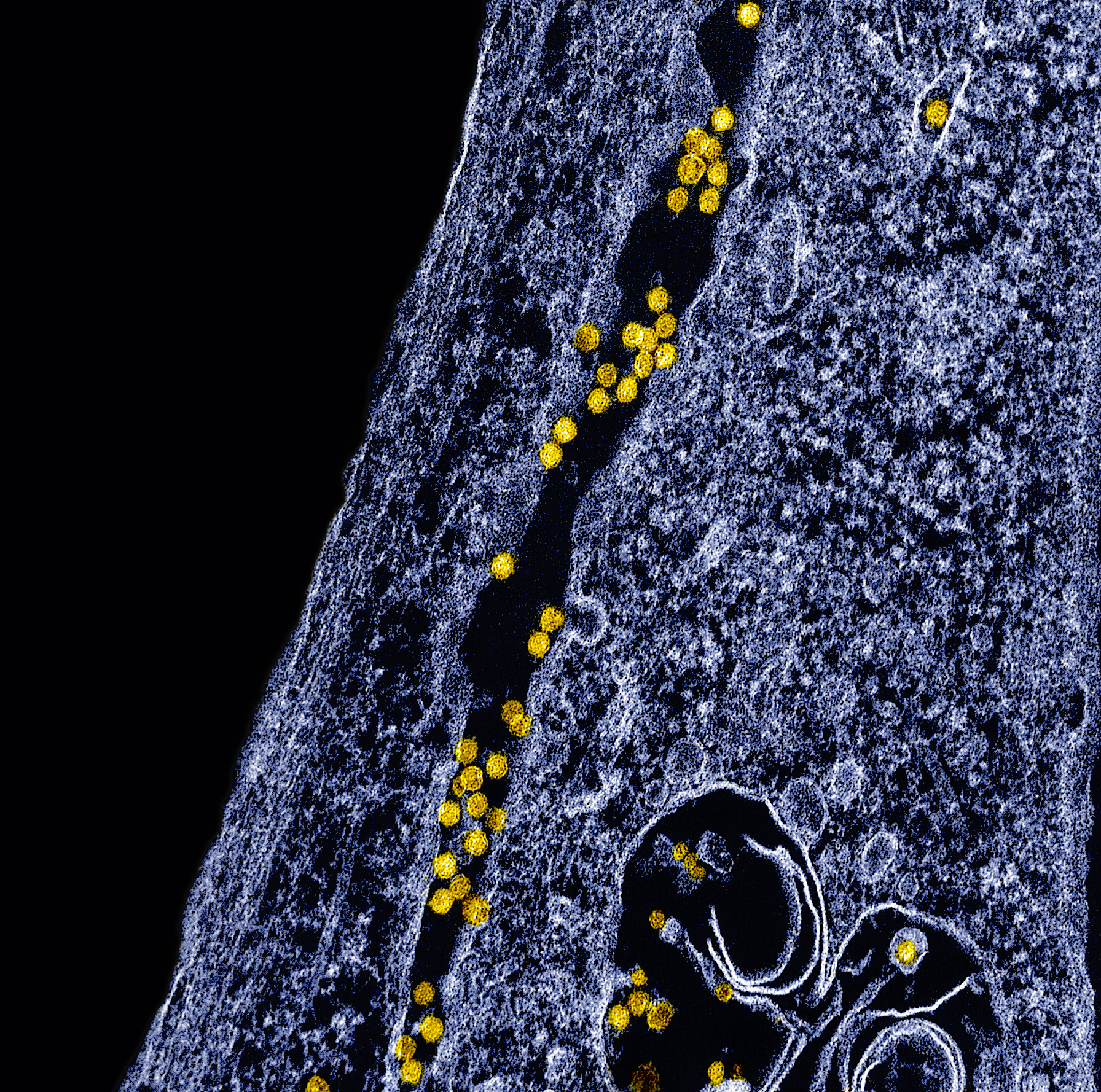
Transmission electron micrograph of West Nile virus particles (yellow) replicating within the cytoplasm of an infected VERO E6 cell (blue). Credit: NIAID
Summary
edit
| Description |
English: Transmission electron micrograph of West Nile virus particles (yellow) replicating within the cytoplasm of an infected VERO E6 cell (blue). Image captured at the NIAID Integrated Research Facility (IRF) in Fort Detrick, Maryland. Credit: NIAID |
| Date | |
| Source | https://www.flickr.com/photos/niaid/53813127810/ |
| Author | NIAID |
Licensing
edit
This file is licensed under the Creative Commons Attribution 2.0 Generic license.
- You are free:
- to share – to copy, distribute and transmit the work
- to remix – to adapt the work
- Under the following conditions:
- attribution – You must give appropriate credit, provide a link to the license, and indicate if changes were made. You may do so in any reasonable manner, but not in any way that suggests the licensor endorses you or your use.
| This image was originally posted to Flickr by NIAID at https://flickr.com/photos/54591706@N02/53813127810. It was reviewed on 24 June 2024 by FlickreviewR 2 and was confirmed to be licensed under the terms of the cc-by-2.0. |
File history
Click on a date/time to view the file as it appeared at that time.
| Date/Time | Thumbnail | Dimensions | User | Comment | |
|---|---|---|---|---|---|
| current | 14:43, 24 June 2024 | | 1,792 × 1,778 (4.14 MB) | Visualspecialist (talk | contribs) | Uploaded a work by NIAID from https://www.flickr.com/photos/niaid/53813127810/ with UploadWizard |
You cannot overwrite this file.
File usage on Commons
There are no pages that use this file.